Switch to Gallery View
Image and Video Gallery
This is a searchable collection of scientific photos, illustrations, and videos. The images and videos in this gallery are licensed under Creative Commons Attribution Non-Commercial ShareAlike 3.0. This license lets you remix, tweak, and build upon this work non-commercially, as long as you credit and license your new creations under identical terms.
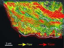
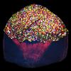
Intracellular forces
2799
Force vectors computed from actin cytoskeleton flow. This is an example of NIH-supported research on single-cell analysis. Gaudenz Danuser, Harvard Medical School View Media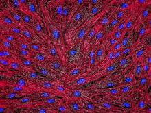
DNA and actin in cultured fibroblast cells
3670
DNA (blue) and actin (red) in cultured fibroblast cells. Tom Deerinck, National Center for Microscopy and Imaging Research (NCMIR) View Media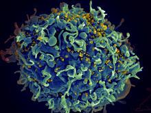
HIV, the AIDS virus, infecting a human cell
3638
This human T cell (blue) is under attack by HIV (yellow), the virus that causes AIDS. Seth Pincus, Elizabeth Fischer, and Austin Athman, National Institute of Allergy and Infectious Diseases, National Institutes of Health View Media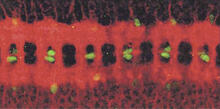
Nerve and glial cells in fruit fly embryo
1091
Glial cells (stained green) in a fruit fly developing embryo have survived thanks to a signaling pathway initiated by neighboring nerve cells (stained red). Hermann Steller, Rockefeller University View Media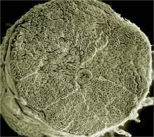
NCMIR human spinal nerve
3387
Spinal nerves are part of the peripheral nervous system. They run within the spinal column to carry nerve signals to and from all parts of the body. Tom Deerinck, National Center for Microscopy and Imaging Research (NCMIR) View Media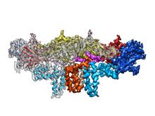
Dengue virus membrane protein structure
3758
Dengue virus is a mosquito-borne illness that infects millions of people in the tropics and subtropics each year. Like many viruses, dengue is enclosed by a protective membrane. Hong Zhou, UCLA View Media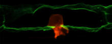
Anchor cell in basement membrane
2707
An anchor cell (red) pushes through the basement membrane (green) that surrounds it. Elliott Hagedorn, Duke University. View Media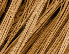
Scanning electron microscopy of collagen fibers
3735
This image shows collagen, a fibrous protein that's the main component of the extracellular matrix (ECM). Collagen is a strong, ropelike molecule that forms stretch-resistant fibers. Tom Deerinck, National Center for Microscopy and Imaging Research (NCMIR) View MediaCone cell
1271
The cone cell of the eye allows you to see in color. Appears in the NIGMS booklet Inside the Cell. Judith Stoffer View Media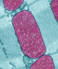
Mitochondria from rat heart muscle cell
3661
These mitochondria (red) are from the heart muscle cell of a rat. Mitochondria have an inner membrane that folds in many places (and that appears here as striations). National Center for Microscopy and Imaging Research View Media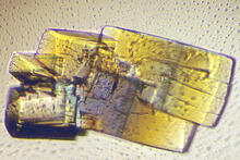
RNase A (2)
2402
A crystal of RNase A protein created for X-ray crystallography, which can reveal detailed, three-dimensional protein structures. Alex McPherson, University of California, Irvine View Media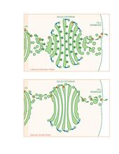
Golgi theories
1278
Two models for how material passes through the Golgi apparatus: the vesicular shuttle model and the cisternae maturation model. Judith Stoffer View Media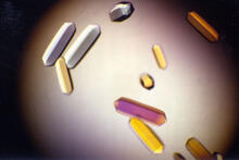
Fungal lipase (1)
2395
Crystals of fungal lipase protein created for X-ray crystallography, which can reveal detailed, three-dimensional protein structures. Alex McPherson, University of California, Irvine View Media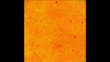
Cell-like compartments emerging from scrambled frog eggs 3
6589
Cell-like compartments spontaneously emerge from scrambled frog eggs. Endoplasmic reticulum (red) and microtubules (green) are visible. Video created using epifluorescence microscopy. Xianrui Cheng, Stanford University School of Medicine. View Media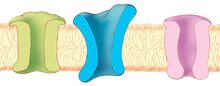
Ion channels
1284
The body uses a variety of ion channels to transport small molecules across cell membranes. Judith Stoffer View Media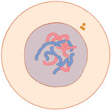
Mitosis - interphase
1316
A cell in interphase, at the start of mitosis: Chromosomes duplicate, and the copies remain attached to each other. Judith Stoffer View Media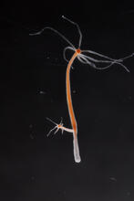
Hydra 04
2440
Hydra magnipapillata is an invertebrate animal used as a model organism to study developmental questions, for example the formation of the body axis. Hiroshi Shimizu, National Institute of Genetics in Mishima, Japan View Media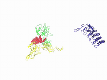
A molecular switch strips transcription factor from DNA
3729
In this video, Rice University scientists used molecular modeling with a mathematical algorithm called AWSEM (for associative memory, water-mediated, structure and energy model) and structural data to Davit Potoyan and Peter Wolynes View Media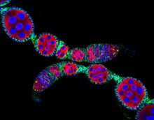
Confocal microscopy image of two Drosophila ovarioles
5772
Ovarioles in female insects are tubes in which egg cells (called oocytes) form at one end and complete their development as they reach the other end of the tube. 2004 Olympus BioScapes Competition View Media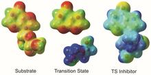
Enzyme transition states
3429
The molecule on the left is an electrostatic potential map of the van der Waals surface of the transition state for human purine nucleoside phosphorylase. Vern Schramm, Albert Einstein College of Medicine of Yeshiva University View Media
Genetically identical mycobacteria respond differently to antibiotic 2
5752
Antibiotic resistance in microbes is a serious health concern. So researchers have turned their attention to how bacteria undo the action of some antibiotics. Bree Aldridge, Tufts University View Media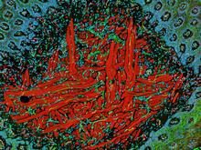
NCMIR Tongue 2
5811
Microscopy image of a tongue. One in a series of two, see image 5810 National Center for Microscopy and Imaging Research (NCMIR) View Media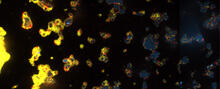
V. Cholerae Biofilm
3580
Industrious V. cholerae bacteria (yellow) tend to thrive in denser biofilms (left) while moochers (red) thrive in weaker biofilms (right). View Media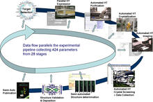
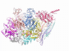
High-throughput protein structure determination pipeline
2364
This slide shows the technologies that the Joint Center for Structural Genomics developed for going from gene to structure and how the technologies have been integrated into a high-throughput pipeline Joint Center for Structural Genomics View Media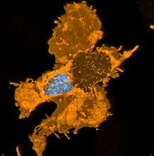
Zebrafish pigment cell
5754
Pigment cells are cells that give skin its color. David Parichy, University of Washington View MediaTracking cells in a gastrulating zebrafish embryo
6776
During development, a zebrafish embryo is transformed from a ball of cells into a recognizable body plan by sweeping convergence and extension cell movements. This process is called gastrulation. Liliana Solnica-Krezel, Washington University School of Medicine in St. Louis. View Media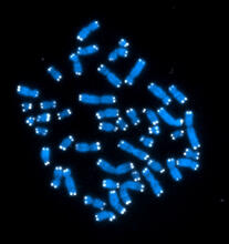
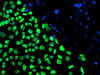
Telomeres
2626
The 46 human chromosomes are shown in blue, with the telomeres appearing as white pinpoints. Hesed Padilla-Nash and Thomas Ried, the National Cancer Institute, a part of NIH View Media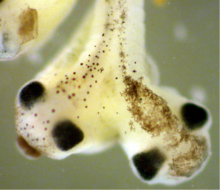
Two-headed Xenopus laevis tadpole
2755
Xenopus laevis, the African clawed frog, has long been used as a research organism for studying embryonic development. Michael Klymkowsky, University of Colorado, Boulder View Media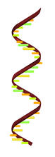
RNA strand
2554
Ribonucleic acid (RNA) has a sugar-phosphate backbone and the bases adenine (A), cytosine (C), guanine (G), and uracil (U). Crabtree + Company View Media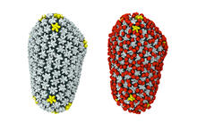
Cryo-EM reveals how the HIV capsid attaches to a human protein to evade immune detection
3755
The illustration shows the capsid of human immunodeficiency virus (HIV) whose molecular features were resolved with cryo-electron microscopy (cryo-EM). Juan R. Perilla, University of Illinois at Urbana-Champaign View Media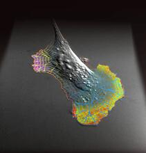
Biosensors illustration
2802
A rendering of an activity biosensor image overlaid with a cell-centered frame of reference used for image analysis of signal transduction. Gaudenz Danuser, Harvard Medical School View Media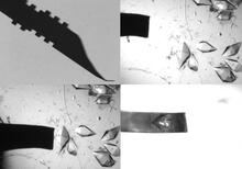
Mounting of protein crystals
2368
Automated methods using micromachined silicon are used at the Northeast Collaboratory for Structural Genomics to mount protein crystals for X-ray crystallography. The Northeast Collaboratory for Structural Genomics View Media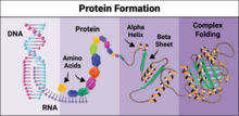
Protein formation
6603
Proteins are 3D structures made up of smaller units. DNA is transcribed to RNA, which in turn is translated into amino acids. NIGMS, with the folded protein illustration adapted from Jane Richardson, Duke University Medical Center View Media
Retinal pigment epithelium derived from human ES cells
3286
This color-enhanced image is a scanning electron microscope image of retinal pigment epithelial (RPE) cells derived from human embryonic stem cells. David Hinton lab, University of Southern California, via CIRM View Media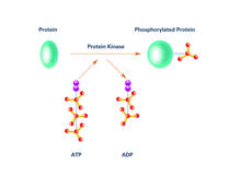
Kinases (with labels)
2535
Kinases are enzymes that add phosphate groups (red-yellow structures) to proteins (green), assigning the proteins a code. Crabtree + Company View Media
TEM cross-section of C. elegans (roundworm)
5759
The worm Caenorhabditis elegans is a popular laboratory animal because its small size and fairly simple body make it easy to study. Piali Sengupta, Brandeis University View Media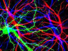
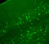
Brain cells in the hippocampus
3688
Hippocampal cells in culture with a neuron in green, showing hundreds of the small protrusions known as dendritic spines. Shelley Halpain, UC San Diego View Media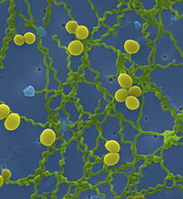
Leptospira bacteria
1166
Leptospira, shown here in green, is a type (genus) of elongated, spiral-shaped bacteria. Infection can cause Weil's disease, a kind of jaundice, in humans. Tina Weatherby Carvalho, University of Hawaii at Manoa View Media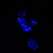
Fluorescence in situ hybridization (FISH) in mouse ES cells shows DNA interactions
3296
Researchers used fluorescence in situ hybridization (FISH) to confirm the presence of long range DNA-DNA interactions in mouse embryonic stem cells. Kathrin Plath, University of California, Los Angeles View Media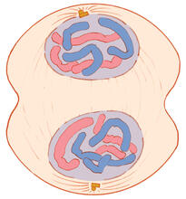
Mitosis - telophase
1332
Telophase during mitosis: Nuclear membranes form around each of the two sets of chromosomes, the chromosomes begin to spread out, and the spindle begins to break down. Judith Stoffer View Media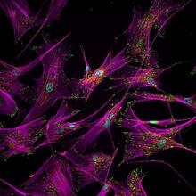
Sticky stem cells
3457
Like a group of barnacles hanging onto a rock, these human cells hang onto a matrix coated glass slide. Ankur Singh and Andrés García, Georgia Institute of Technology View Media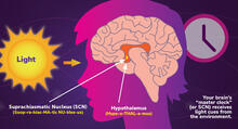
Circadian rhythms and the SCN
6613
Circadian rhythms are physical, mental, and behavioral changes that follow a 24-hour cycle. NIGMS View Media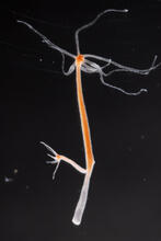
Hydra 02
2438
Hydra magnipapillata is an invertebrate animal used as a model organism to study developmental questions, for example the formation of the body axis. Hiroshi Shimizu, National Institute of Genetics in Mishima, Japan View Media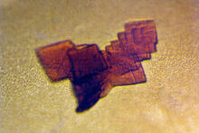
Sheep hemoglobin crystal
2392
A crystal of sheep hemoglobin protein created for X-ray crystallography, which can reveal detailed, three-dimensional protein structures. Alex McPherson, University of California, Irvine View Media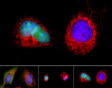
Apoptosis reversed
3486
Two healthy cells (bottom, left) enter into apoptosis (bottom, center) but spring back to life after a fatal toxin is removed (bottom, right; top). Hogan Tang of the Denise Montell Lab, Johns Hopkins University School of Medicine View Media
Biopixels
3266
Bioengineers were able to coax bacteria to blink in unison on microfluidic chips. This image shows a small chip with about 500 blinking bacterial colonies or biopixels. Jeff Hasty Lab, UC San Diego View Media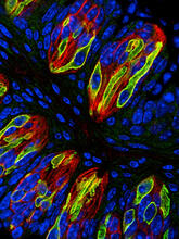
Taste buds signal different tastes through ATP release
3444
Taste buds in a mouse tongue epithelium with types I, II, and III taste cells visualized by cell-type-specific fluorescent antibodies. Aki Taruno, Perelman School of Medicine, University of Pennsylvania View Media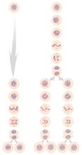
Mitosis and meiosis compared
1333
Meiosis is used to make sperm and egg cells. During meiosis, a cell's chromosomes are copied once, but the cell divides twice. Judith Stoffer View Media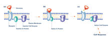
G switch (with labels and stages)
2538
The G switch allows our bodies to respond rapidly to hormones. G proteins act like relay batons to pass messages from circulating hormones into cells. Crabtree + Company View Media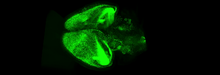
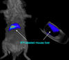
Mouse brain 3
6931
Various views of a mouse brain that was genetically modified so that subpopulations of its neurons glow. Prayag Murawala, MDI Biological Laboratory and Hannover Medical School. View Media