Switch to Gallery View
Image and Video Gallery
This is a searchable collection of scientific photos, illustrations, and videos. The images and videos in this gallery are licensed under Creative Commons Attribution Non-Commercial ShareAlike 3.0. This license lets you remix, tweak, and build upon this work non-commercially, as long as you credit and license your new creations under identical terms.
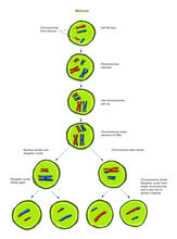
Meiosis illustration (with labels)
2546
Meiosis is the process whereby a cell reduces its chromosomes from diploid to haploid in creating eggs or sperm. Crabtree + Company View Media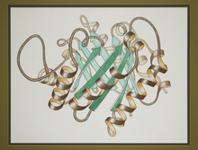
Early ribbon drawing of a protein
2748
This ribbon drawing of a protein hand drawn and colored by researcher Jane Richardson in 1981 helped originate the ribbon representation of proteins that is now ubiquitous in molecular graphics. Jane Richardson, Duke University Medical Center View Media
Mitosis - interphase
1316
A cell in interphase, at the start of mitosis: Chromosomes duplicate, and the copies remain attached to each other. Judith Stoffer View Media
Migrating pigment cells
5758
Pigment cells are cells that give skin its color. David Parichy, University of Washington View Media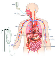
A drug's life in the body (with labels)
2528
A drug's life in the body. Medicines taken by mouth (oral) pass through the liver before they are absorbed into the bloodstream. Crabtree + Company View Media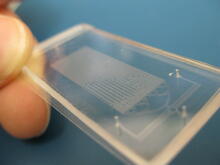
Microfluidic chip
3265
Microfluidic chips have many uses in biology labs. Jeff Hasty Lab, UC San Diego View Media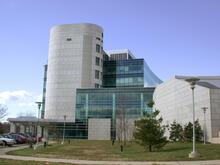
Natcher Building 04
1084
NIGMS staff are located in the Natcher Building on the NIH campus. Alisa Machalek, National Institute of General Medical Sciences View Media
DDR2 Receptors Attach to Collagen in Breast Tumor
3478
On the left, the boundary of a breast tumor (yellow) attaches to collagen fibers that are closest to it (green) using DDR2. On the right, a tumor without DDR2 remains disconnected from the collagen. Callie Corsa and Suzanne Ponik, Washington University School of Medicine in St. Louis View Media
Mouse heart muscle cells 02
3283
This image shows neonatal mouse heart cells. These cells were grown in the lab on a chip that aligns the cells in a way that mimics what is normally seen in the body. Kara McCloskey lab, University of California, Merced, via CIRM View Media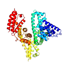
Serum albumin structure 2
3745
Serum albumin (SA) is the most abundant protein in the blood plasma of mammals. SA has a characteristic heart-shape structure and is a highly versatile protein. Wladek Minor, University of Virginia View Media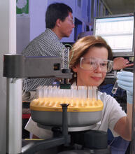
Protein purification robot
2375
Irina Dementieva, a biochemist, and Youngchang Kim, a biophysicist and crystallographer, work with the first robot of its type in the U.S. to automate protein purification. Midwest Center for Structural Genomics View Media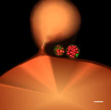
Multivesicular bodies containing intralumenal vesicles assemble at the vacuole 1
5769
Collecting and transporting cellular waste and sorting it into recylable and nonrecylable pieces is a complex business in the cell. Matthew West and Greg Odorizzi, University of Colorado View Media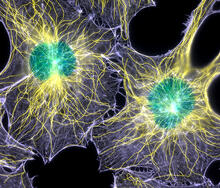
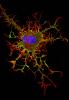
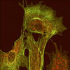
Colorful cells
2428
Actin (purple), microtubules (yellow), and nuclei (green) are labeled in these cells by immunofluorescence. This image won first place in the Nikon 2003 Small World photo competition. Torsten Wittmann, Scripps Research Institute View Media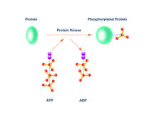
Kinases (with labels)
2535
Kinases are enzymes that add phosphate groups (red-yellow structures) to proteins (green), assigning the proteins a code. Crabtree + Company View Media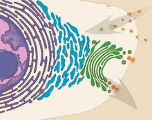
Vesicle traffic
1283
This illustration shows vesicle traffic inside a cell. Judith Stoffer View Media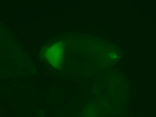
Fly cells live
2315
If a picture is worth a thousand words, what's a movie worth? Denise Montell, Johns Hopkins University School of Medicine View Media
Natcher Building 02
1082
NIGMS staff are located in the Natcher Building on the NIH campus. Alisa Machalek, National Institute of General Medical Sciences View Media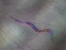
C. elegans trapped by carnivorous fungus
6963
Real-time footage of Caenorhabditis elegans, a tiny roundworm, trapped by a carnivorous fungus, Arthrobotrys dactyloides. Michael Shribak, Marine Biological Laboratory/University of Chicago. View Media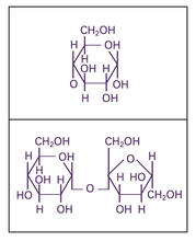
Glucose and sucrose
2500
Glucose (top) and sucrose (bottom) are sugars made of carbon, hydrogen, and oxygen atoms. Carbohydrates include simple sugars like these and are the main source of energy for the human body. Crabtree + Company View Media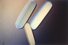
Fungal lipase (2)
2411
Crystals of fungal lipase protein created for X-ray crystallography, which can reveal detailed, three-dimensional protein structures. Alex McPherson, University of California, Irvine View Media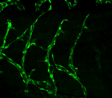
Disrupted vascular development in frog embryos
3403
Disassembly of vasculature in kdr:GFP frogs following addition of 250 µM TBZ. Related to images 3404 and 3505. Hye Ji Cha, University of Texas at Austin View Media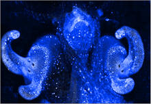
Pores on the surface of the Hawaiian bobtail squid light organ
7016
The light organ (~0.5 mm across) of a juvenile Hawaiian bobtail squid, Euprymna scolopes, stained blue. Margaret J. McFall-Ngai, Carnegie Institution for Science/California Institute of Technology, and Edward G. Ruby, California Institute of Technology. View Media
Golgi
1275
The Golgi complex, also called the Golgi apparatus or, simply, the Golgi. Judith Stoffer View Media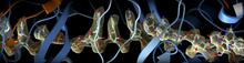
CRISPR
6351
RNA incorporated into the CRISPR surveillance complex is positioned to scan across foreign DNA. Cryo-EM density from a 3Å reconstruction is shown as a yellow mesh. NRAMM National Resource for Automated Molecular Microscopy http://nramm.nysbc.org/nramm-images/ Source: Bridget Carragher View Media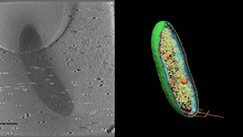
Cryo-electron tomography of a Caulobacter bacterium
6569
3D image of Caulobacter bacterium with various components highlighted: cell membranes (red and blue), protein shell (green), protein factories known as ribosomes (yellow), and storage granules Peter Dahlberg, Stanford University. View Media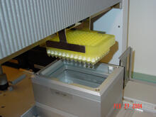
Protein purification robot in action 01
2369
A robot is transferring 96 purification columns to a vacuum manifold for subsequent purification procedures. The Northeast Collaboratory for Structural Genomics View Media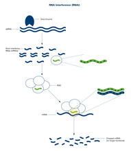
RNA interference (with labels)
2559
RNA interference or RNAi is a gene-silencing process in which double-stranded RNAs trigger the destruction of specific RNAs. Crabtree + Company View Media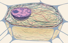
Cytoskeleton
1272
The three fibers of the cytoskeleton--microtubules in blue, intermediate filaments in red, and actin in green--play countless roles in the cell. Judith Stoffer View MediaCentral dogma, illustrated (with labels and numbers for stages)
2549
DNA encodes RNA, which encodes protein. DNA is transcribed to make messenger RNA (mRNA). The mRNA sequence (dark red strand) is complementary to the DNA sequence (blue strand). Crabtree + Company View Media
Natcher Building 07
1087
NIGMS staff are located in the Natcher Building on the NIH campus. Alisa Machalek, National Institute of General Medical Sciences View Media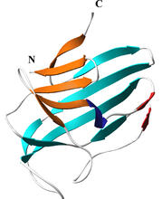
Secreted protein from Mycobacteria
2379
Model of a major secreted protein of unknown function, which is only found in mycobacteria, the class of bacteria that causes tuberculosis. Mycobacterium Tuberculosis Center, PSI View Media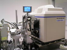
Chromium X-ray source
2361
In the determination of protein structures by X-ray crystallography, this unique soft (l = 2.29Å) X-ray source is used to collect anomalous scattering data from protein crystals containing light atoms The Southeast Collaboratory for Structural Genomics View Media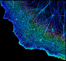
3D image of actin in a cell
3749
Actin is an essential protein in a cell's skeleton (cytoskeleton). It forms a dense network of thin filaments in the cell. Xiaowei Zhuang, Howard Hughes Medical Institute, Harvard University View Media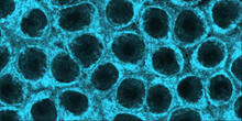
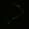
Movements of myosin
2324
Inside the fertilized egg cell of a fruit fly, we see a type of myosin (related to the protein that helps muscles contract) made to glow by attaching a fluorescent protein. Victoria Foe, University of Washington View Media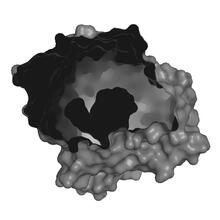
X-ray co-crystal structure of Src kinase bound to a DNA-templated macrocycle inhibitor 2
3414
X-ray co-crystal structure of Src kinase bound to a DNA-templated macrocycle inhibitor. Markus A. Seeliger, Stony Brook University Medical School and David R. Liu, Harvard University View Media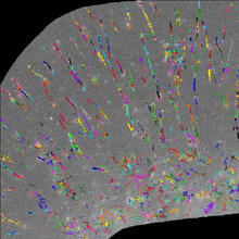
Trajectories of labeled cell receptors
2801
Trajectories of single molecule labeled cell surface receptors. This is an example of NIH-supported research on single-cell analysis. Gaudenz Danuser, Harvard Medical School View Media
Xenopus laevis egg
2753
Xenopus laevis, the African clawed frog, has long been used as a model organism for studying embryonic development. Michael Klymkowsky, University of Colorado, Boulder View Media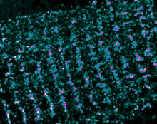
Single-Molecule Imaging
3339
This is a super-resolution light microscope image taken by Hiro Hakozaki and Masa Hoshijima of NCMIR. Tom Deerinck, NCMIR View Media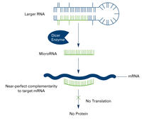
Dicer generates microRNAs (with labels)
2557
The enzyme Dicer generates microRNAs by chopping larger RNA molecules into tiny Velcro®-like pieces. MicroRNAs stick to mRNA molecules and prevent the mRNAs from being made into proteins. Crabtree + Company View MediaAssembly of the HIV capsid
5729
The HIV capsid is a pear-shaped structure that is made of proteins the virus needs to mature and become infective. John Grime and Gregory Voth, The University of Chicago View Media
Pulsating response to stress in bacteria - video
3254
By attaching fluorescent proteins to the genetic circuit responsible for B. subtilis's stress response, researchers can observe the cells' pulses as green flashes. Michael Elowitz, Caltech University View Media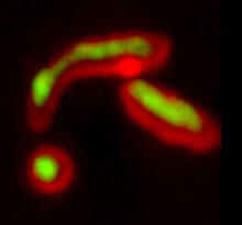
Misfolded proteins within in the mitochondria
5878
Misfolded proteins (green) within mitochondria (red). Related to video 5877. Rong Li rong@jhu.edu Department of Chemical and Biomolecular Engineering, Whiting School of Engineering, Johns Hopkins University, Baltimore, Maryland 21218, USA. View Media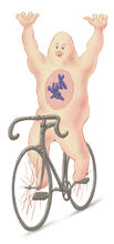
Bicycling cell
1337
A humorous treatment of the concept of a cycling cell. Judith Stoffer View Media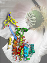
Atomic Structure of Poppy Enzyme
3422
The atomic structure of the morphine biosynthetic enzyme salutaridine reductase bound to the cofactor NADPH. The substrate salutaridine is shown entering the active site. Judy Coyle, Donald Danforth Plant Science Center View Media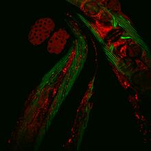
Closeup of fluorescent C. elegans showing muscle and ribosomal protein
6583
Closeup of C. elegans, tiny roundworms, with a ribosomal protein glowing red and muscle fibers glowing green. Researchers used these worms to study a molecular pathway that affects aging. Jarod Rollins, Mount Desert Island Biological Laboratory. View Media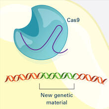
CRISPR Illustration Frame 4
6488
This illustration shows, in simplified terms, how the CRISPR-Cas9 system can be used as a gene-editing tool. National Institute of General Medical Sciences. View Media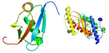
Ubiquitin-fold modifier 1 from C. elegans
2388
Solution NMR structure of protein target WR41 (left) from C. elegans. Northeast Structural Genomics Consortium View Media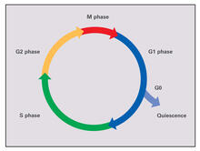
Cell cycle (with labels)
2499
Cells progress through a cycle that consists of phases for growth (G1, S, and G2) and division (M). Cells become quiescent when they exit this cycle (G0). Crabtree + Company View Media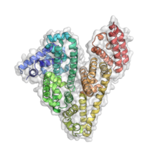
Serum albumin structure 3
3746
Serum albumin (SA) is the most abundant protein in the blood plasma of mammals. SA has a characteristic heart-shape structure and is a highly versatile protein. Wladek Minor, University of Virginia View Media