Switch to Gallery View
Image and Video Gallery
This is a searchable collection of scientific photos, illustrations, and videos. The images and videos in this gallery are licensed under Creative Commons Attribution Non-Commercial ShareAlike 3.0. This license lets you remix, tweak, and build upon this work non-commercially, as long as you credit and license your new creations under identical terms.
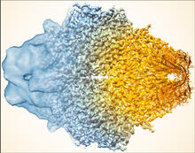
Beta-galactosidase montage showing cryo-EM improvement--gradient background
5883
Composite image of beta-galactosidase showing how cryo-EM’s resolution has improved dramatically in recent years. Older images to the left, more recent to the right. Veronica Falconieri, Sriram Subramaniam Lab, National Cancer Institute View Media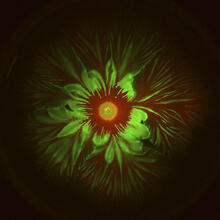
Floral pattern in a mixture of two bacterial species, Acinetobacter baylyi and Escherichia coli, grown on a semi-solid agar for 48 hours (photo 2)
6555
Floral pattern emerging as two bacterial species, motile Acinetobacter baylyi (red) and non-motile Escherichia coli (green), are grown together for 48 hours on 1% agar surface from a sma L. Xiong et al, eLife 2020;9: e48885 View Media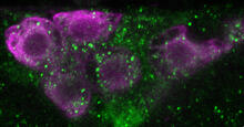
Insulin production and fat sensing in fruit flies
6982
Fourteen neurons (magenta) in the adult Drosophila brain produce insulin, and fat tissue sends packets of lipids to the brain via the lipoprotein carriers (green). Akhila Rajan, Fred Hutchinson Cancer Center View Media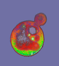
Cells frozen in time
2307
The fledgling field of X-ray microscopy lets researchers look inside whole cells rapidly frozen to capture their actions at that very moment. Here, a yeast cell buds before dividing into two. Carolyn Larabell, University of California, San Francisco, and the Lawrence Berkeley National Laboratory View Media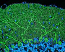
Mouse cerebellum
5795
The cerebellum is the brain's locomotion control center. Found at the base of your brain, the cerebellum is a single layer of tissue with deep folds like an accordion. National Center for Microscopy and Imaging Research (NCMIR) View Media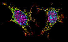
Two mouse fibroblast cells
6789
Two mouse fibroblasts, one of the most common types of cells in mammalian connective tissue. They play a key role in wound healing and tissue repair. Dylan T. Burnette, Vanderbilt University School of Medicine. View Media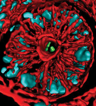
Mouse colon with gut bacteria
3566
A section of mouse colon with gut bacteria (center, in green) residing within a protective pocket. Sarkis K. Mazmanian, California Institute of Technology View Media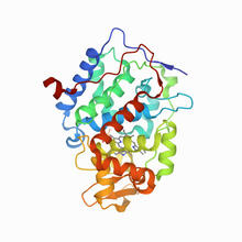
CCP enzyme
6762
The enzyme CCP is found in the mitochondria of baker’s yeast. Scientists study the chemical reactions that CCP triggers, which involve a water molecule, iron, and oxygen. Protein Data Bank. View Media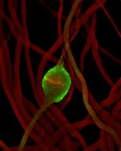
Fruit fly sperm cells
2433
Developing fruit fly spermatids require caspase activity (green) for the elimination of unwanted organelles and cytoplasm via apoptosis. Hermann Steller, Rockefeller University View Media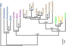
Dinosaur evolutionary tree
2474
Analysis of 68 million-year-old collagen molecule fragments preserved in a T. Chris Organ, Harvard University View Media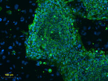
Human embryonic stem cells on feeder cells
3274
This fluorescent microscope image shows human embryonic stem cells whose nuclei are stained green. Blue staining shows the surrounding supportive feeder cells. Michael Longaker lab, Stanford University School of Medicine, via CIRM View Media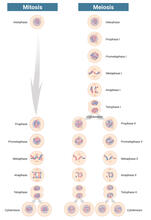
Mitosis and meiosis compared-labeled
6788
Meiosis is used to make sperm and egg cells. During meiosis, a cell's chromosomes are copied once, but the cell divides twice. Judith Stoffer View Media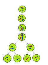
Meiosis illustration
2545
Meiosis is the process whereby a cell reduces its chromosomes from diploid to haploid in creating eggs or sperm. Crabtree + Company View Media
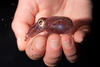
Adult Hawaiian bobtail squid burying in the sand
7012
Each morning, the nocturnal Hawaiian bobtail squid, Euprymna scolopes, hides from predators by digging into the sand. At dusk, it leaves the sand again to hunt. Margaret J. McFall-Ngai, Carnegie Institution for Science/California Institute of Technology, and Edward G. Ruby, California Institute of Technology. View Media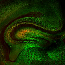
Mouse Brain Cross Section
5886
The brain sections are treated with fluorescent antibodies specific to a particular protein and visualized using serial electron microscopy (SEM). Anton Maximov, The Scripps Research Institute, La Jolla, CA View Media
Cytoscape network wiring diagram 2
2749
This image integrates the thousands of known molecular and genetic interactions happening inside our bodies using a computer program called Cytoscape. Trey Ideker, University of California, San Diego View Media
Tongue 1
5810
Microscopy image of tongue. One in a series of two, see image 5811 National Center for Microscopy and Imaging Research (NCMIR) View Media
Group of Culex quinquefasciatus mosquito larvae
6770
Mosquito larvae with genes edited by CRISPR. Valentino Gantz, University of California, San Diego. View Media
Bacillus anthracis being killed
3525
Bacillus anthracis (anthrax) cells being killed by a fluorescent trans-translation inhibitor, which disrupts bacterial protein synthesis. Kenneth Keiler, Penn State University View Media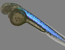
Zebrafish embryo
6897
A zebrafish embryo showing its natural colors. Zebrafish have see-through eggs and embryos, making them ideal research organisms for studying the earliest stages of development. Michael Shribak, Marine Biological Laboratory/University of Chicago. View Media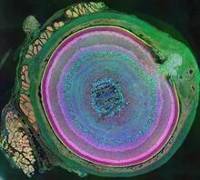
A mammalian eye has approximately 70 different cell types
3641
The incredible complexity of a mammalian eye (in this case from a mouse) is captured here. Each color represents a different type of cell. Bryan William Jones and Robert E. Marc, University of Utah View Media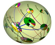
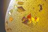
Statistical cartography
2331
Like a world of its own, this sphere represents all the known chemical reactions in the E. coli bacterium. Luis A. Nunes Amaral, Northwestern University View Media
Lysosomes
1282
Lysosomes have powerful enzymes and acids to digest and recycle cell materials. Judith Stoffer View Media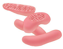
Mitochondria
1287
Bean-shaped mitochondria are cells' power plants. These organelles have their own DNA and replicate independently. The highly folded inner membranes are the site of energy generation. Judith Stoffer View Media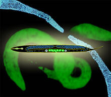
Worms and human infertility
2333
This montage of tiny, transparent C. elegans--or roundworms--may offer insight into understanding human infertility. Abby Dernburg, Lawrence Berkeley National Laboratory View Media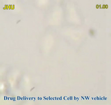
Precisely Delivering Chemical Cargo to Cells
3779
Moving protein or other molecules to specific cells to treat or examine them has been a major biological challenge. Nature Nanotechnology View Media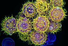
Pollen grains: male germ cells in plants and a cause of seasonal allergies
3609
Those of us who get sneezy and itchy-eyed every spring or fall may have pollen grains, like those shown here, to blame. Edna, Gil, and Amit Cukierman, Fox Chase Cancer Center, Philadelphia, Pa. View Media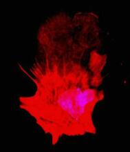
Polarized cells- 01
3332
Cells move forward with lamellipodia and filopodia supported by networks and bundles of actin filaments. Proper, controlled cell movement is a complex process. Rong Li and Praveen Suraneni, Stowers Institute for Medical Research View Media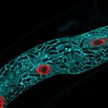
Microsporidia in roundworm 2
5778
Many disease-causing microbes manipulate their host’s metabolism and cells for their own ends. Keir Balla and Emily Troemel, University of California San Diego View Media
A bundle of myelinated peripheral nerve cells (axons)
3737
The extracellular matrix (ECM) is most prevalent in connective tissues but also is present between the stems (axons) of nerve cells. Tom Deerinck, National Center for Microscopy and Imaging Research (NCMIR) View Media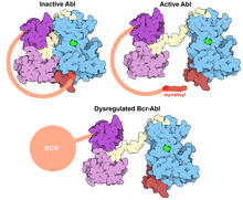
Protein kinases as cancer chemotherapy targets
7004
Protein kinases—enzymes that add phosphate groups to molecules—are cancer chemotherapy targets because they play significant roles in almost all aspects of cell function, are tightly regulated, and co Amy Wu and Christine Zardecki, RCSB Protein Data Bank. View Media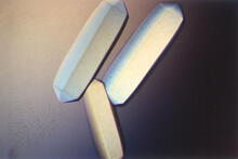
Fungal lipase (2)
2411
Crystals of fungal lipase protein created for X-ray crystallography, which can reveal detailed, three-dimensional protein structures. Alex McPherson, University of California, Irvine View Media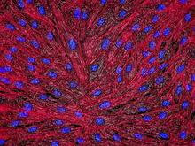
DNA and actin in cultured fibroblast cells
3670
DNA (blue) and actin (red) in cultured fibroblast cells. Tom Deerinck, National Center for Microscopy and Imaging Research (NCMIR) View Media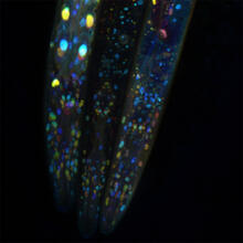
Mosaicism in C. elegans (Black Background)
6532
In the worm C. elegans, double-stranded RNA made in neurons can silence matching genes in a variety of cell types through the transport of RNA between cells. Snusha Ravikumar, Ph.D., University of Maryland, College Park, and Antony M. Jose, Ph.D., University of Maryland, College Park View Media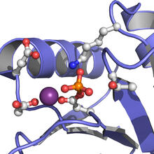
Active Site of E. coli response regulator PhoB
3412
Active site of E. coli response regulator PhoB. Ann Stock, Rutgers University View Media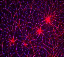
In vitro assembly of a cell-signaling pathway
3787
T cells are white blood cells that are important in defending the body against bacteria, viruses and other pathogens. Xiaolei Su, HHMI Whitman Center of the Marine Biological Laboratory View Media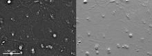
Epithelial cell migration
6899
High-resolution time lapse of epithelial (skin) cell migration and wound healing. It shows an image taken every 13 seconds over the course of almost 14 minutes. Michael Shribak, Marine Biological Laboratory/University of Chicago. View Media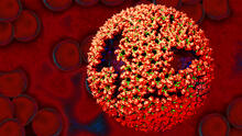
Molecular model of freshly made Rous sarcoma virus (RSV)
3771
Viruses have been the foes of animals and other organisms for time immemorial. Boon Chong Goh, University of Illinois at Urbana-Champaign View Media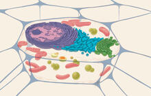
Animal cell
1274
A typical animal cell, sliced open to reveal a cross-section of organelles. Judith Stoffer View Media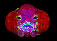
Zebrafish larva
5881
You are face to face with a 6-day-old zebrafish larva. What look like eyes will become nostrils, and the bulges on either side will become eyes. Oscar Ruiz and George Eisenhoffer, University of Texas MD Anderson Cancer Center, Houston View Media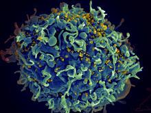
HIV, the AIDS virus, infecting a human cell
3638
This human T cell (blue) is under attack by HIV (yellow), the virus that causes AIDS. Seth Pincus, Elizabeth Fischer, and Austin Athman, National Institute of Allergy and Infectious Diseases, National Institutes of Health View Media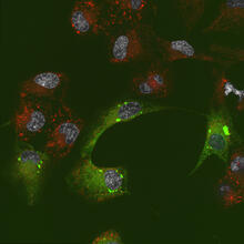
Endoplasmic reticulum abnormalities 2
6774
Human cells with the gene that codes for the protein FIT2 deleted. After an experimental intervention, they are expressing a nonfunctional version of FIT2, shown in green. Michel Becuwe, Harvard University. View Media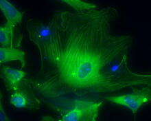
Smooth muscle from human ES cells
3288
These smooth muscle cells were derived from human embryonic stem cells. The nuclei are stained blue, and the proteins of the cytoskeleton are stained green. Alexey Terskikh lab, Burnham Institute for Medical Research, via CIRM View Media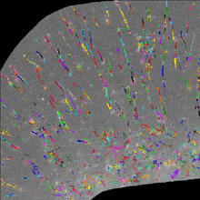
Trajectories of labeled cell receptors
2801
Trajectories of single molecule labeled cell surface receptors. This is an example of NIH-supported research on single-cell analysis. Gaudenz Danuser, Harvard Medical School View Media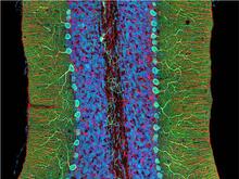
Mouse cerebellum close-up
3371
The cerebellum is the brain's locomotion control center. Every time you shoot a basketball, tie your shoe or chop an onion, your cerebellum fires into action. National Center for Microscopy and Imaging Research (NCMIR) View Media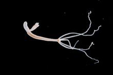
Hydra 05
2441
Hydra magnipapillata is an invertebrate animal used as a model organism to study developmental questions, for example the formation of the body axis. Hiroshi Shimizu, National Institute of Genetics in Mishima, Japan View Media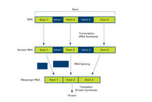
Introns (with labels)
2551
Genes are often interrupted by stretches of DNA (introns, blue) that do not contain instructions for making a protein. Crabtree + Company View Media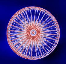
Arachnoidiscus diatom
6902
An Arachnoidiscus diatom with a diameter of 190µm. Michael Shribak, Marine Biological Laboratory/University of Chicago. View Media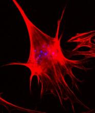
Polarized cells- 02
3333
Cells move forward with lamellipodia and filopodia supported by networks and bundles of actin filaments. Proper, controlled cell movement is a complex process. Rong Li and Praveen Suraneni, Stowers Institute for Medical Research View Media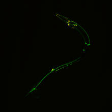
Neural circuits in worms similar to those in humans
3252
Green and yellow fluorescence mark the processes and cell bodies of some C. elegans neurons. Shawn Xu, University of Michigan View Media