Switch to Gallery View
Image and Video Gallery
This is a searchable collection of scientific photos, illustrations, and videos. The images and videos in this gallery are licensed under Creative Commons Attribution Non-Commercial ShareAlike 3.0. This license lets you remix, tweak, and build upon this work non-commercially, as long as you credit and license your new creations under identical terms.
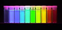
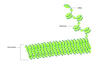
Nano-rainbow
2326
These vials may look like they're filled with colored water, but they really contain nanocrystals reflecting different colors under ultraviolet light. Shuming Nie, Emory University View Media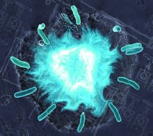
Supernova bacteria
2725
Bacteria engineered to act as genetic clocks flash in synchrony. Here, a "supernova" burst in a colony of coupled genetic clocks just after reaching critical cell density. Jeff Hasty, UCSD View Media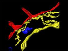
Computer model of cell membrane
2636
A computer model of the cell membrane, where the plasma membrane is red, endoplasmic reticulum is yellow, and mitochondria are blue. Bridget Wilson, University of New Mexico View Media
An adult Hawaiian bobtail squid
7013
An adult female Hawaiian bobtail squid, Euprymna scolopes, with its mantle cavity exposed from the underside. Margaret J. McFall-Ngai, Carnegie Institution for Science/California Institute of Technology, and Edward G. Ruby, California Institute of Technology. View Media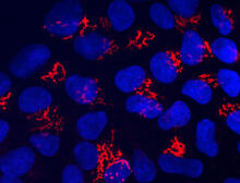
Suicidal Stem Cells
3341
Embryonic stem cells store pre-activated Bax (red) in the Golgi, near the nucleus (blue). Featured in the June 21, 2012, issue of Biomedical Beat. Mohanish Deshmukh View Media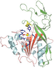
Human aspartoacylase
2352
Model of aspartoacylase, a human enzyme involved in brain metabolism. Center for Eukaryotic Structural Genomics, PSI View Media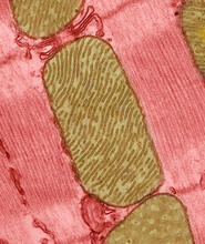
Mitochondria from rat heart muscle cell_2
3664
These mitochondria (brown) are from the heart muscle cell of a rat. Mitochondria have an inner membrane that folds in many places (and that appears here as striations). National Center for Microscopy and Imaging Research View Media
Leading cells with light
2708
A blue laser beam turns on a protein that helps this human cancer cell move. Responding to the stimulus, the protein, called Rac1, first creates ruffles at the edge of the cell. Yi Wu, University of North Carolina View Media
CRISPR Illustration Frame 3
6487
This illustration shows, in simplified terms, how the CRISPR-Cas9 system can be used as a gene-editing tool. National Institute of General Medical Sciences. View Media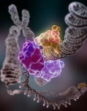
Repairing DNA
3493
Like a watch wrapped around a wrist, a special enzyme encircles the double helix to repair a broken strand of DNA. Tom Ellenberger, Washington University School of Medicine View Media
Smooth ER
1292
The endoplasmic reticulum comes in two types: Rough ER is covered with ribosomes and prepares newly made proteins; smooth ER specializes in making lipids and breaking down toxic molecules. Judith Stoffer View Media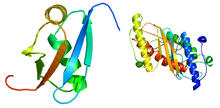
Ubiquitin-fold modifier 1 from C. elegans
2388
Solution NMR structure of protein target WR41 (left) from C. elegans. Northeast Structural Genomics Consortium View Media
A drug's life in the body (with labels)
2528
A drug's life in the body. Medicines taken by mouth (oral) pass through the liver before they are absorbed into the bloodstream. Crabtree + Company View Media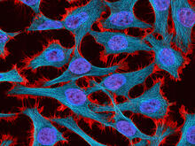
HeLa cells
3521
Multiphoton fluorescence image of HeLa cells stained with the actin binding toxin phalloidin (red), microtubules (cyan) and cell nuclei (blue). Nikon RTS2000MP custom laser scanning microscope. National Center for Microscopy and Imaging Research (NCMIR) View Media
Breast cancer cells change migration phenotypes
6986
Cancer cells can change their migration phenotype, which includes their shape and the way that they move to invade different tissues. Bo Sun, Oregon State University. View Media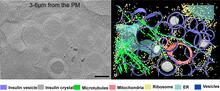
Cryo-ET cell cross-section visualizing insulin vesicles
6607
On the left, a cross-section slice of a rat pancreas cell captured using cryo-electron tomography (cryo-ET). On the right, a color-coded, 3D version of the image highlighting cell structures. Xianjun Zhang, University of Southern California. View Media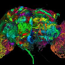
Color coding of the Drosophila brain - black background
5868
This image results from a research project to visualize which regions of the adult fruit fly (Drosophila) brain derive from each neural stem cell. Yong Wan from Charles Hansen’s lab, University of Utah. Data preparation and visualization by Masayoshi Ito in the lab of Kei Ito, University of Tokyo. View Media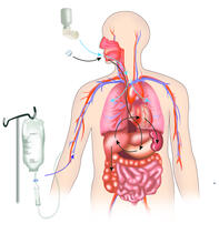
A drug's life in the body
2527
A drug's life in the body. Medicines taken by mouth pass through the liver before they are absorbed into the bloodstream. Crabtree + Company View Media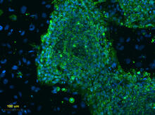
Human embryonic stem cells on feeder cells
3274
This fluorescent microscope image shows human embryonic stem cells whose nuclei are stained green. Blue staining shows the surrounding supportive feeder cells. Michael Longaker lab, Stanford University School of Medicine, via CIRM View Media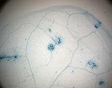
Disease-resistant Arabidopsis leaf
2781
This is a magnified view of an Arabidopsis thaliana leaf a few days after being exposed to the pathogen Hyaloperonospora arabidopsidis. Jeff Dangl, University of North Carolina, Chapel Hill View Media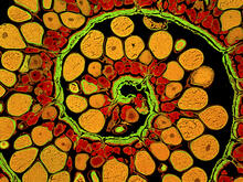
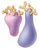
Anglerfish ovary cross-section
3620
This image captures the spiral-shaped ovary of an anglerfish in cross-section. Once matured, these eggs will be released in a gelatinous, floating mass. James E. Hayden, The Wistar Institute, Philadelphia, Pa. View Media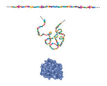
Building blocks and folding of proteins
2508
Proteins are made of amino acids hooked end-to-end like beads on a necklace. To become active, proteins must twist and fold into their final, or "native," conformation. Crabtree + Company View Media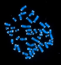
Telomeres
2626
The 46 human chromosomes are shown in blue, with the telomeres appearing as white pinpoints. Hesed Padilla-Nash and Thomas Ried, the National Cancer Institute, a part of NIH View Media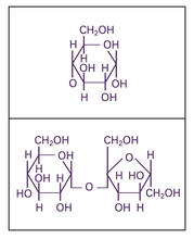
Glucose and sucrose
2500
Glucose (top) and sucrose (bottom) are sugars made of carbon, hydrogen, and oxygen atoms. Carbohydrates include simple sugars like these and are the main source of energy for the human body. Crabtree + Company View Media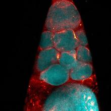
Fruit fly nurse cells during egg development
6753
In many animals, the egg cell develops alongside sister cells. Adam C. Martin, Massachusetts Institute of Technology. View Media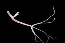
Hydra 06
2442
Hydra magnipapillata is an invertebrate animal used as a model organism to study developmental questions, for example the formation of the body axis. Hiroshi Shimizu, National Institute of Genetics in Mishima, Japan View Media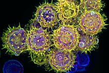
Pollen grains: male germ cells in plants and a cause of seasonal allergies
3609
Those of us who get sneezy and itchy-eyed every spring or fall may have pollen grains, like those shown here, to blame. Edna, Gil, and Amit Cukierman, Fox Chase Cancer Center, Philadelphia, Pa. View Media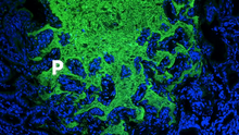
Pathways: What is Basic Science?
6539
Learn about basic science, sometimes called “pure” or “fundamental” science, and how it contributes to the development of medical treatments. National Institute of General Medical Sciences View Media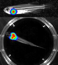
Bioluminescent imaging in adult zebrafish - lateral and overhead view
3556
Luciferase-based imaging enables visualization and quantification of internal organs and transplanted cells in live adult zebrafish. Kenneth Poss, Duke University View Media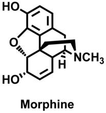
Morphine Structure
3438
The chemical structure of the morphine molecule Judy Coyle, Donald Danforth Plant Science Center View Media
Biofilm formed by a pathogen
6518
A biofilm is a highly organized community of microorganisms that develops naturally on certain surfaces. Scott Chimileski, Ph.D., and Roberto Kolter, Ph.D., Harvard Medical School. View Media
Katanin protein regulates anaphase
2594
The microtubule severing protein, katanin, localizes to chromosomes and regulates anaphase A in mitosis. David Sharp, Albert Einstein College of Medicine View Media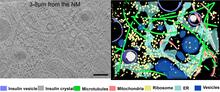
Cryo-ET cross-section of a rat pancreas cell
6608
On the left, a cross-section slice of a rat pancreas cell captured using cryo-electron tomography (cryo-ET). On the right, a 3D, color-coded version of the image highlighting cell structures. Xianjun Zhang, University of Southern California. View Media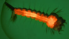
Culex quinquefasciatus mosquito larva
6769
A mosquito larva with genes edited by CRISPR. The red-orange glow is a fluorescent protein used to track the edits. Valentino Gantz, University of California, San Diego. View Media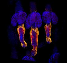
Growing hair follicle stem cells
3499
Wound healing requires the action of stem cells. Hermann Steller, Rockefeller University View Media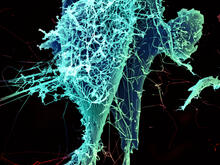
String-like Ebola virus peeling off an infected cell
3619
After multiplying inside a host cell, the stringlike Ebola virus is emerging to infect more cells. Heinz Feldmann, Peter Jahrling, Elizabeth Fischer and Anita Mora, National Institute of Allergy and Infectious Diseases, National Institutes of Health View Media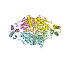
Thymidylate synthase complementing protein from Thermotoga maritime
2387
A model of thymidylate synthase complementing protein from Thermotoga maritime. Joint Center for Structural Genomics, PSI View Media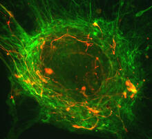
Human ES cells differentiating into neurons
3276
This image shows hundreds of human embryonic stem cells in various stages of differentiating into neurons. Guoping Fan lab, University of California, Los Angeles, via CIRM View Media
Mapping metabolic activity
2319
Like a map showing heavily traveled roads, this mathematical model of metabolic activity inside an E. coli cell shows the busiest pathway in white. Albert-László Barabási, University of Notre Dame View Media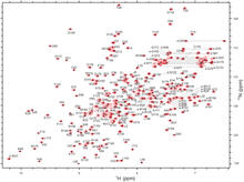
2-D NMR
2299
A two-dimensional NMR spectrum of a protein, in this case a 2D 1H-15N HSQC NMR spectrum of a 228 amino acid DNA/RNA-binding protein. Dr. Xiaolian Gao's laboratory at the University of Houston View Media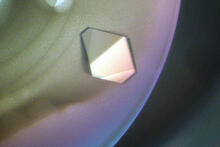
Bacterial glucose isomerase
2409
A crystal of bacterial glucose isomerase protein created for X-ray crystallography, which can reveal detailed, three-dimensional protein structures. Alex McPherson, University of California, Irvine View Media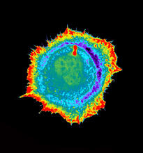
Seeing signaling protein activation in cells 01
2451
Cdc42, a member of the Rho family of small guanosine triphosphatase (GTPase) proteins, regulates multiple cell functions, including motility, proliferation, apoptosis, and cell morphology. Klaus Hahn, University of North Carolina, Chapel Hill Medical School View Media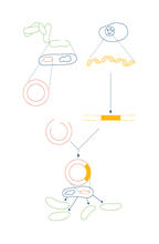
Recombinant DNA
2564
To splice a human gene into a plasmid, scientists take the plasmid out of an E. coli bacterium, cut the plasmid with a restriction enzyme, and splice in human DNA. Crabtree + Company View Media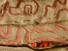
Scanning electron microscopy of the ECM on the surface of a calf muscle
3739
This image shows the extracellular matrix (ECM) on the surface of a soleus (lower calf) muscle in light brown and blood vessels in pink. Tom Deerinck, National Center for Microscopy and Imaging Research (NCMIR) View Media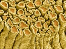
Myelinated axons 1
3396
Myelinated axons in a rat spinal root. Tom Deerinck, National Center for Microscopy and Imaging Research (NCMIR) View Media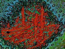
NCMIR Tongue 2
5811
Microscopy image of a tongue. One in a series of two, see image 5810 National Center for Microscopy and Imaging Research (NCMIR) View Media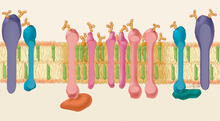
Lipid raft
1285
Researchers have learned much of what they know about membranes by constructing artificial membranes in the laboratory. Judith Stoffer View Media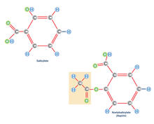
Aspirin (with labels)
2530
Acetylsalicylate (bottom) is the aspirin of today. Crabtree + Company View Media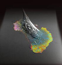
Biosensors illustration
2802
A rendering of an activity biosensor image overlaid with a cell-centered frame of reference used for image analysis of signal transduction. Gaudenz Danuser, Harvard Medical School View Media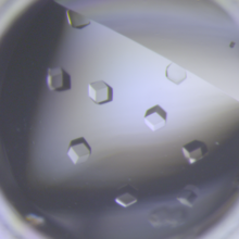
Crystals of CCD-1 in complex with cefotaxime
6764
CCD-1 is an enzyme produced by the bacterium Clostridioides difficile that helps it resist antibiotics. Keith Hodgson, Stanford University. View Media