Switch to Gallery View
Image and Video Gallery
This is a searchable collection of scientific photos, illustrations, and videos. The images and videos in this gallery are licensed under Creative Commons Attribution Non-Commercial ShareAlike 3.0. This license lets you remix, tweak, and build upon this work non-commercially, as long as you credit and license your new creations under identical terms.
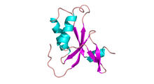
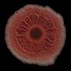
Antitoxin GhoS (Illustration 1)
3427
Structure of the bacterial antitoxin protein GhoS. GhoS inhibits the production of a bacterial toxin, GhoT, which can contribute to antibiotic resistance. Rebecca Page and Wolfgang Peti, Brown University and Thomas K. Wood, Pennsylvania State University View Media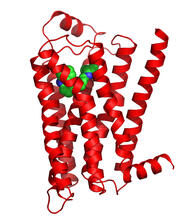
Beta 2-adrenergic receptor
3358
The receptor is shown bound to a partial inverse agonist, carazolol. Raymond Stevens, The Scripps Research Institute View Media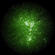
Cytoscape network wiring diagram 2
2749
This image integrates the thousands of known molecular and genetic interactions happening inside our bodies using a computer program called Cytoscape. Trey Ideker, University of California, San Diego View Media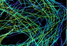
Microtubules in African green monkey cells
6891
Microtubules in African green monkey cells. Microtubules are strong, hollow fibers that provide cells with structural support. Melike Lakadamyali, Perelman School of Medicine at the University of Pennsylvania. View Media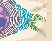
Vesicle traffic
1283
This illustration shows vesicle traffic inside a cell. Judith Stoffer View Media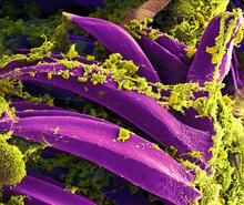
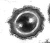
Bubonic plague bacteria on part of the digestive system in a rat flea
3576
Here, bubonic plague bacteria (yellow) are shown in the digestive system of a rat flea (purple). The bubonic plague killed a third of Europeans in the mid-14th century. NIAID View Media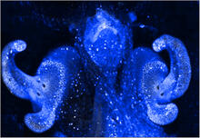
Pores on the surface of the Hawaiian bobtail squid light organ
7016
The light organ (~0.5 mm across) of a juvenile Hawaiian bobtail squid, Euprymna scolopes, stained blue. Margaret J. McFall-Ngai, Carnegie Institution for Science/California Institute of Technology, and Edward G. Ruby, California Institute of Technology. View Media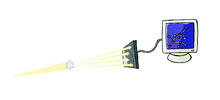
X-ray crystallography
2511
X-ray crystallography allows researchers to see structures too small to be seen by even the most powerful microscopes. Crabtree + Company View Media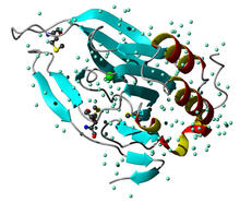
Cysteine dioxygenase from mouse
2347
Model of the mammalian iron enzyme cysteine dioxygenase from a mouse. Center for Eukaryotic Structural Genomics, PSI View Media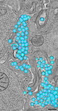
HIV-1 virus in the colon
3571
A tomographic reconstruction of the colon shows the location of large pools of HIV-1 virus particles (in blue) located in the spaces between adjacent cells. Mark Ladinsky, California Institute of Technology View Media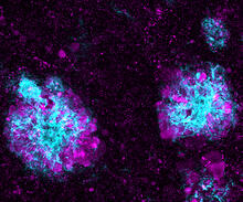
Lysosome clusters around amyloid plaques
5771
It's probably most people's least favorite activity, but we still need to do it--take out our trash. Otherwise our homes will get cluttered and smelly, and eventually, we'll get sick. Swetha Gowrishankar and Shawn Ferguson, Yale School of Medicine View Media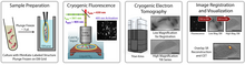
Correlative imaging by annotation with single molecules (CIASM) process
6568
These images illustrate a technique combining cryo-electron tomography and super-resolution fluorescence microscopy called correlative imaging by annotation with single molecules (CIASM). Peter Dahlberg, Stanford University. View Media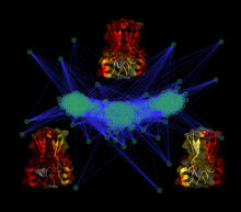
Cluster analysis of mysterious protein
3295
Researchers use cluster analysis to study protein shape and function. Each green circle represents one potential shape of the protein mitoNEET. Patricia Jennings and Elizabeth Baxter, University of California, San Diego View Media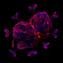
Wild-type and mutant fruit fly ovaries
6806
The two large, central, round shapes are ovaries from a typical fruit fly (Drosophila melanogaster). Vladimir I. Gelfand, Feinberg School of Medicine, Northwestern University. View Media
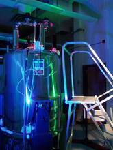
Superconducting magnet
1120
Superconducting magnet for NMR research, from the February 2003 profile of Dorothee Kern in Findings. Mike Lovett View Media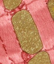
Mitochondria from rat heart muscle cell_2
3664
These mitochondria (brown) are from the heart muscle cell of a rat. Mitochondria have an inner membrane that folds in many places (and that appears here as striations). National Center for Microscopy and Imaging Research View Media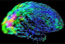
Mapping brain differences
2419
This image of the human brain uses colors and shapes to show neurological differences between two people. Arthur Toga, University of California, Los Angeles View Media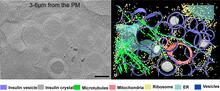
Cryo-ET cell cross-section visualizing insulin vesicles
6607
On the left, a cross-section slice of a rat pancreas cell captured using cryo-electron tomography (cryo-ET). On the right, a color-coded, 3D version of the image highlighting cell structures. Xianjun Zhang, University of Southern California. View Media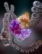
Repairing DNA
3493
Like a watch wrapped around a wrist, a special enzyme encircles the double helix to repair a broken strand of DNA. Tom Ellenberger, Washington University School of Medicine View Media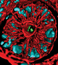
Mouse colon with gut bacteria
3566
A section of mouse colon with gut bacteria (center, in green) residing within a protective pocket. Sarkis K. Mazmanian, California Institute of Technology View Media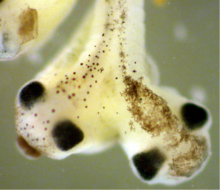
Two-headed Xenopus laevis tadpole
2755
Xenopus laevis, the African clawed frog, has long been used as a research organism for studying embryonic development. Michael Klymkowsky, University of Colorado, Boulder View Media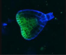
Early development in Arabidopsis
2733
Early on, this Arabidopsis plant embryo picks sides: While one end will form the shoot, the other will take root underground. Zachery R. Smith, Jeff Long lab at the Salk Institute for Biological Studies View Media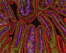
NCMIR Intestine-2
3390
The small intestine is where most of our nutrients from the food we eat are absorbed into the bloodstream. Tom Deerinck, National Center for Microscopy and Imaging Research (NCMIR) View Media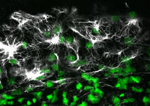
Vimentin in a quail embryo
2807
Confocal image showing high levels of the protein vimentin (white) at the edge zone of a quail embryo. Cell nuclei are labeled green. Andrés Garcia, Georgia Tech View Media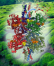
Structure of Glutamate Dehydrogenase
3421
Some children are born with a mutation in a regulatory site on this enzyme that causes them to over-secrete insulin when they consume protein. Judy Coyle, Donald Danforth Plant Science Center View Media
Mapping human genetic variation
2443
This map paints a colorful portrait of human genetic variation around the world. Noah Rosenberg and Martin Soave, University of Michigan View Media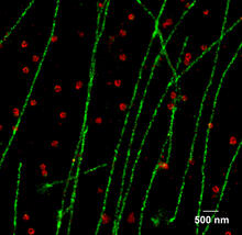
Multicolor STORM
2325
In 2006, scientists developed an optical microscopy technique enabling them to clearly see individual molecules within cells. In 2007, they took the technique, abbreviated STORM, a step further. Xiaowei Zhuang, Harvard University View Media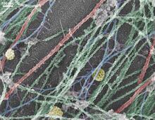
Cells use bubble-like structures called vesicles to transport cargo
3634
Cells use bubble-like structures called vesicles (yellow) to import, transport, and export cargo and in cellular communication. A single cell may be filled with thousands of moving vesicles.Tatyana Svitkina, University of Pennsylvania View Media
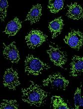
Calcium uptake during ATP production in mitochondria
3449
Living primary mouse embryonic fibroblasts. Mitochondria (green) stained with the mitochondrial membrane potential indicator, rhodamine 123. Nuclei (blue) are stained with DAPI. Lili Guo, Perelman School of Medicine, University of Pennsylvania View Media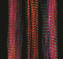
Three muscle fibers; the middle has a defect found in some neuromuscular diseases
3630
Of the three muscle fibers shown here, the one on the right and the one on the left are normal. The middle fiber is deficient a large protein called nebulin (blue). Christopher Pappas and Carol Gregorio, University of Arizona View Media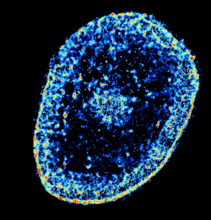
Chromatin in human tenocyte
6893
The nucleus of a degenerating human tendon cell, also known as a tenocyte. It has been color-coded based on the density of chromatin—a substance made up of DNA and proteins. Melike Lakadamyali, Perelman School of Medicine at the University of Pennsylvania. View Media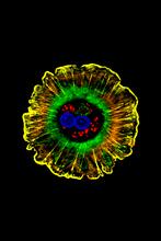
Human liver cell (hepatocyte)
3610
Hepatocytes, like the one shown here, are the most abundant type of cell in the human liver. Donna Beer Stolz, University of Pittsburgh View Media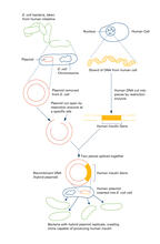
Recombinant DNA (with labels)
2565
To splice a human gene (in this case, the one for insulin) into a plasmid, scientists take the plasmid out of an E. Crabtree + Company View Media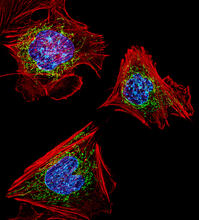
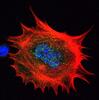
Fibroblasts with nuclei in blue, energy factories in green and the actin cytoskeleton in red
3624
The cells shown here are fibroblasts, one of the most common cells in mammalian connective tissue. These particular cells were taken from a mouse embryo. Dylan Burnette, NICHD View Media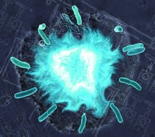
Supernova bacteria
2725
Bacteria engineered to act as genetic clocks flash in synchrony. Here, a "supernova" burst in a colony of coupled genetic clocks just after reaching critical cell density. Jeff Hasty, UCSD View Media
Natcher Building 05
1085
NIGMS staff are located in the Natcher Building on the NIH campus. Alisa Machalek, National Institute of General Medical Sciences View Media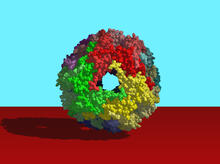
Heat shock protein complex from Methanococcus jannaschii
2385
Model based on X-ray crystallography of the structure of a small heat shock protein complex from the bacteria, Methanococcus jannaschii. Berkeley Structural Genomics Center, PSI-1 View Media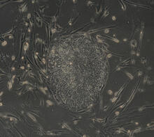
Induced stem cells from adult skin 04
2606
The human skin cells pictured contain genetic modifications that make them pluripotent, essentially equivalent to embryonic stem cells. James Thomson, University of Wisconsin-Madison View Media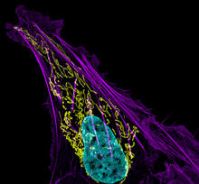
Bone cancer cell
3626
This image shows an osteosarcoma cell with DNA in blue, energy factories (mitochondria) in yellow, and actin filaments—part of the cellular skeleton—in purple. Dylan Burnette and Jennifer Lippincott-Schwartz, NICHD View Media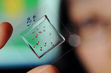
Automated Worm Sorter - 4
3475
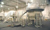
Georgia Tech associate professor Hang Lu holds a microfluidic chip that is part of a system that uses artificial intelligence and cutting-edge image processing to automatically examine large number of Georgia Tech/Gary Meek View Media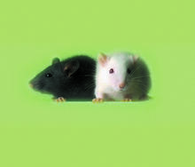
Lab mice
1069
Many researchers use the mouse (Mus musculus) as a model organism to study mammalian biology. Bill Branson, National Institutes of Health View Media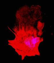
Polarized cells- 01
3332
Cells move forward with lamellipodia and filopodia supported by networks and bundles of actin filaments. Proper, controlled cell movement is a complex process. Rong Li and Praveen Suraneni, Stowers Institute for Medical Research View Media
Natcher Building 06
1086
NIGMS staff are located in the Natcher Building on the NIH campus. Alisa Machalek, National Institute of General Medical Sciences View Media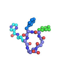
X-ray co-crystal structure of Src kinase bound to a DNA-templated macrocycle inhibitor 7
3419
X-ray co-crystal structure of Src kinase bound to a DNA-templated macrocycle inhibitor. Markus A. Seeliger, Stony Brook University Medical School and David R. Liu, Harvard University View Media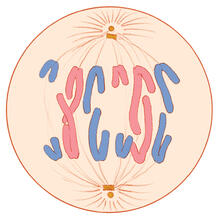
Mitosis - anaphase
1328
A cell in anaphase during mitosis: Chromosomes separate into two genetically identical groups and move to opposite ends of the spindle. Judith Stoffer View Media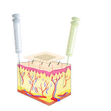
Drugs enter skin
2531
Drugs enter different layers of skin via intramuscular, subcutaneous, or transdermal delivery methods. See image 2532 for a labeled version of this illustration. Crabtree + Company View Media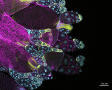
Fruit fly ovary
6522
In this image of a stained fruit fly ovary, the ovary is packed with immature eggs (with DNA stained blue). The cytoskeleton (in pink) is a collection of fibers that gives a cell shape and support. Crystal D. Rogers, Ph.D., University of California, Davis, School of Veterinary Medicine; and Mariano A. Loza-Coll, Ph.D., California State University, Northridge. View Media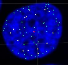
Telomeres on outer edge of nucleus during cell division
3484
New research shows telomeres moving to the outer edge of the nucleus after cell division, suggesting these caps that protect chromosomes also may play a role in organizing DNA. Laure Crabbe, Jamie Kasuboski and James Fitzpatrick, Salk Institute for Biological Studies View Media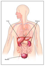
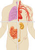
Body toxins (with labels)
2497
Body organs such as the liver and kidneys process chemicals and toxins. These "target" organs are susceptible to damage caused by these substances. Crabtree + Company View Media