Switch to Gallery View
Image and Video Gallery
This is a searchable collection of scientific photos, illustrations, and videos. The images and videos in this gallery are licensed under Creative Commons Attribution Non-Commercial ShareAlike 3.0. This license lets you remix, tweak, and build upon this work non-commercially, as long as you credit and license your new creations under identical terms.
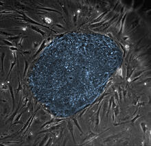
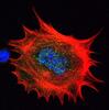
Human embryonic stem cells
2608
The center cluster of cells, colored blue, shows a colony of human embryonic stem cells. James Thomson, University of Wisconsin-Madison View Media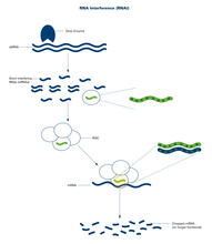
RNA interference (with labels)
2559
RNA interference or RNAi is a gene-silencing process in which double-stranded RNAs trigger the destruction of specific RNAs. Crabtree + Company View Media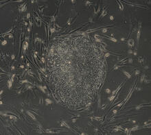
Induced stem cells from adult skin 04
2606
The human skin cells pictured contain genetic modifications that make them pluripotent, essentially equivalent to embryonic stem cells. James Thomson, University of Wisconsin-Madison View Media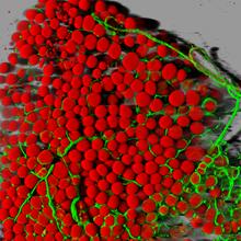
Fat cells (red) and blood vessels (green)
3600
A mouse's fat cells (red) are shown surrounded by a network of blood vessels (green). Daniela Malide, National Heart, Lung, and Blood Institute, National Institutes of Health View Media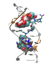
Myotonic dystrophy type 2 genetic defect
3573
Scientists revealed a detailed image of the genetic defect that causes myotonic dystrophy type 2 and used that information to design drug candidates to counteract the disease. Matthew Disney, Scripps Research Institute and Ilyas Yildirim, Northwestern University View Media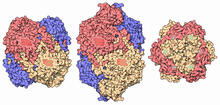
Catalase diversity
7003
Catalases are some of the most efficient enzymes found in cells. Amy Wu and Christine Zardecki, RCSB Protein Data Bank. View Media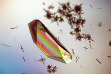
Hen egg lysozyme (1)
2396
Crystals of hen egg lysozyme protein created for X-ray crystallography, which can reveal detailed, three-dimensional protein structures. Alex McPherson, University of California, Irvine View Media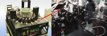
Capillary protein crystallization robot
2357
This ACAPELLA robot for capillary protein crystallization grows protein crystals, freezes them, and centers them without manual intervention. Structural Genomics of Pathogenic Protozoa Consortium View Media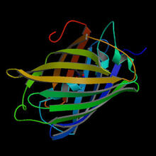
Fruitful dyes
2317
These colorful, computer-generated ribbons show the backbone of a molecule that glows a fluorescent red. Roger Y. Tsien, University of California, San Diego View Media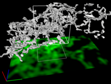
Dense tubular matrices in the peripheral endoplasmic reticulum (ER) 2
5856
Three-dimensional reconstruction of a tubular matrix in a thin section of the peripheral endoplasmic reticulum between the plasma membranes of the cell. Jennifer Lippincott-Schwartz, Howard Hughes Medical Institute Janelia Research Campus, Virginia View Media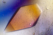
Pig trypsin (1)
2400
A crystal of porcine trypsin protein created for X-ray crystallography, which can reveal detailed, three-dimensional protein structures. Alex McPherson, University of California, Irvine View Media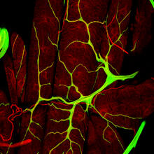
An insect tracheal cell delivers air to muscles
3615
Insects like the fruit fly use an elaborate network of branching tubes called trachea (green) to transport oxygen throughout their bodies. Jayan Nair and Maria Leptin, European Molecular Biology Laboratory, Heidelberg, Germany View Media
Natcher Building 08
1088
NIGMS staff are located in the Natcher Building on the NIH campus. Alisa Machalek, National Institute of General Medical Sciences View Media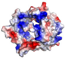
VDAC-1 (3)
2494
The structure of the pore-forming protein VDAC-1 from humans. Gerhard Wagner, Harvard Medical School View Media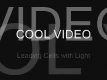
Leading cells with light
2708
A blue laser beam turns on a protein that helps this human cancer cell move. Responding to the stimulus, the protein, called Rac1, first creates ruffles at the edge of the cell. Yi Wu, University of North Carolina View Media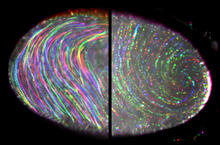
Fruit fly egg ooplasmic streaming
6809
Two fruit fly (Drosophila melanogaster) egg cells, one on each side of the central black line. Vladimir I. Gelfand, Feinberg School of Medicine, Northwestern University. View Media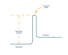
Activation energy (with labels)
2526
To become products, reactants must overcome an energy hill. See image 2525 for an unlabeled version of this illustration. Crabtree + Company View Media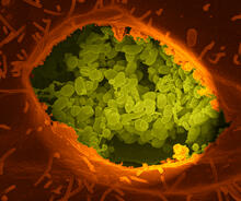
Q fever bacteria in an infected cell
3621
This image shows Q fever bacteria (yellow), which infect cows, sheep, and goats around the world and can infect humans, as well. When caught early, Q fever can be cured with antibiotics. Robert Heinzen, Elizabeth Fischer, and Anita Mora, National Institute of Allergy and Infectious Diseases, National Institutes of Health View Media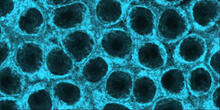
Movements of myosin
2324
Inside the fertilized egg cell of a fruit fly, we see a type of myosin (related to the protein that helps muscles contract) made to glow by attaching a fluorescent protein. Victoria Foe, University of Washington View Media
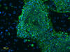
Planarian stem cell colony
3306
Planarians are freshwater flatworms that have powerful abilities to regenerate their bodies, which would seem to make them natural model organisms in which to study stem cells. Peter Reddien, Whitehead Institute View Media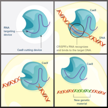
CRISPR Illustration
7036
This illustration shows, in simplified terms, how the CRISPR-Cas9 system can be used as a gene-editing tool. National Institute of General Medical Sciences. View Media
Human skeletal muscle
3677
Cross section of human skeletal muscle. Image taken with a confocal fluorescent light microscope. Tom Deerinck, National Center for Microscopy and Imaging Research (NCMIR) View Media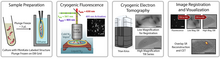
Correlative imaging by annotation with single molecules (CIASM) process
6568
These images illustrate a technique combining cryo-electron tomography and super-resolution fluorescence microscopy called correlative imaging by annotation with single molecules (CIASM). Peter Dahlberg, Stanford University. View Media
Kupffer cell residing in the liver
6535
Kupffer cells appear in the liver during the early stages of mammalian development and stay put throughout life to protect liver cells, clean up old red blood cells, and regulate iron levels. Thomas Deerinck, National Center for Microscopy and Imaging Research, University of California, San Diego. View Media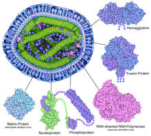
Measles virus proteins
6996
A cross section of the measles virus in which six proteins (enlarged on the outside of the virus) work together to infect cells. Amy Wu and Christine Zardecki, RCSB Protein Data Bank. View Media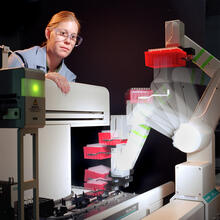
Student overseeing protein cloning robot
2356
Student Christina Hueneke of the Midwest Center for Structural Genomics is overseeing a protein cloning robot. Midwest Center for Structural Genomics View Media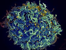
HIV, the AIDS virus, infecting a human cell
3638
This human T cell (blue) is under attack by HIV (yellow), the virus that causes AIDS. Seth Pincus, Elizabeth Fischer, and Austin Athman, National Institute of Allergy and Infectious Diseases, National Institutes of Health View Media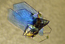
DNase
2410
Crystals of DNase protein created for X-ray crystallography, which can reveal detailed, three-dimensional protein structures. Alex McPherson, University of California, Irvine View Media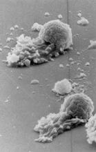
Worm sperm
3489
To develop a system for studying cell motility in unnatrual conditions -- a microscope slide instead of the body -- Tom Roberts and Katsuya Shimabukuro at Florida State University disassembled and rec Tom Roberts, Florida State University View Media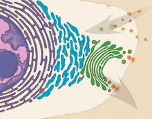
Vesicle traffic
1283
This illustration shows vesicle traffic inside a cell. Judith Stoffer View Media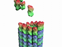
How a microtubule builds and deconstructs
3650
A microtubule, part of the cell's skeleton, builds and deconstructs. View Media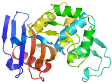
Ribbon diagram of a cefotaxime-CCD-1 complex
6766
CCD-1 is an enzyme produced by the bacterium Clostridioides difficile that helps it resist antibiotics. Keith Hodgson, Stanford University. View Media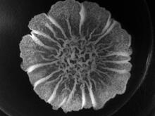
A Bacillus subtilis biofilm grown in a Petri dish
3718
Bacterial biofilms are tightly knit communities of bacterial cells growing on, for example, solid surfaces, such as in water pipes or on teeth. Gürol Süel, UCSD View Media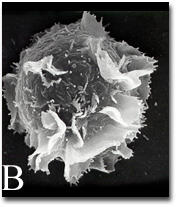
Activated mast cell surface
2637
A scanning electron microscope image of an activated mast cell. This image illustrates the interesting topography of the cell membrane, which is populated with receptors. Bridget Wilson, University of New Mexico View Media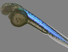
Zebrafish embryo
6897
A zebrafish embryo showing its natural colors. Zebrafish have see-through eggs and embryos, making them ideal research organisms for studying the earliest stages of development. Michael Shribak, Marine Biological Laboratory/University of Chicago. View Media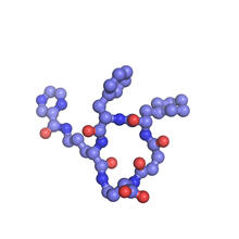
X-ray co-crystal structure of Src kinase bound to a DNA-templated macrocycle inhibitor 6
3418
X-ray co-crystal structure of Src kinase bound to a DNA-templated macrocycle inhibitor. Markus A. Seeliger, Stony Brook University Medical School and David R. Liu, Harvard University View Media
Fruit fly retina 02
2434
Section of a fruit fly retina showing the light-sensing molecules rhodopsin-5 (blue) and rhodopsin-6 (red). Hermann Steller, Rockefeller University View Media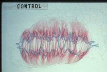
Lily mitosis 09
1022
A light microscope image of a cell from the endosperm of an African globe lily (Scadoxus katherinae). This is one frame of a time-lapse sequence that shows cell division in action. Andrew S. Bajer, University of Oregon, Eugene View Media
The Proteasome: The Cell's Trash Processor in Action
3772
Our cells are constantly removing and recycling molecular waste. This video shows one way cells process their trash. View Media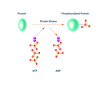
Kinases (with labels)
2535
Kinases are enzymes that add phosphate groups (red-yellow structures) to proteins (green), assigning the proteins a code. Crabtree + Company View Media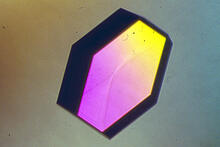
Hen egg lysozyme (2)
2406
A crystal of hen egg lysozyme protein created for X-ray crystallography, which can reveal detailed, three-dimensional protein structures. Alex McPherson, University of California, Irvine View Media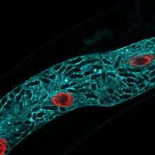
Microsporidia in roundworm 2
5778
Many disease-causing microbes manipulate their host’s metabolism and cells for their own ends. Keir Balla and Emily Troemel, University of California San Diego View Media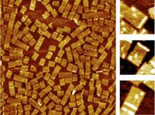
Golden gene chips
2455
A team of chemists and physicists used nanotechnology and DNA's ability to self-assemble with matching RNA to create a new kind of chip for measuring gene activity. Hao Yan and Yonggang Ke, Arizona State University View Media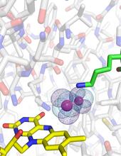
O2 reacting with a flavin-dependent enzyme
3411
Department of Biological Chemistry, University of Michigan View Media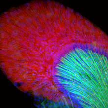
Developing zebrafish fin
3598
Originally from the waters of India, Nepal, and neighboring countries, zebrafish can now be found swimming in science labs (and home aquariums) throughout the world. Jessica Plavicki View Media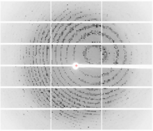
X-ray diffraction pattern from a crystallized cefotaxime-CCD-1 complex
6765
CCD-1 is an enzyme produced by the bacterium Clostridioides difficile that helps it resist antibiotics. Keith Hodgson, Stanford University. View Media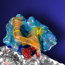
Kinesin moves cellular cargo
3491
A protein called kinesin (blue) is in charge of moving cargo around inside cells and helping them divide. Charles Sindelar, Yale University View Media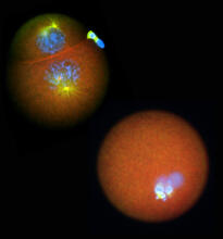
Nucleolinus
2762
The nucleolinus is a cellular compartment that has been a lonely bystander in scientific endeavors. Mary Anne Alliegro, Marine Biological Laboratory View Media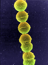
Streptococcus bacteria
1157
Image of Streptococcus, a type (genus) of spherical bacteria that can colonize the throat and back of the mouth. Stroptococci often occur in pairs or in chains, as shown here. Tina Weatherby Carvalho, University of Hawaii at Manoa View Media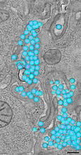
HIV-1 virus in the colon
3571
A tomographic reconstruction of the colon shows the location of large pools of HIV-1 virus particles (in blue) located in the spaces between adjacent cells. Mark Ladinsky, California Institute of Technology View Media