Switch to Gallery View
Image and Video Gallery
This is a searchable collection of scientific photos, illustrations, and videos. The images and videos in this gallery are licensed under Creative Commons Attribution Non-Commercial ShareAlike 3.0. This license lets you remix, tweak, and build upon this work non-commercially, as long as you credit and license your new creations under identical terms.
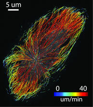
Microtubule growth
2800
Map of microtubule growth rates. Rates are color coded. This is an example of NIH-supported research on single-cell analysis. Gaudenz Danuser, Harvard Medical School View Media
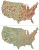
Haplotypes (with labels)
2567
Haplotypes are combinations of gene variants that are likely to be inherited together within the same chromosomal region. Crabtree + Company View Media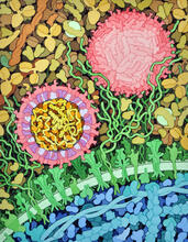
Zika virus
6998
Zika virus is shown in cross section at center left. On the outside, it includes envelope protein (red) and membrane protein (magenta) embedded in a lipid membrane (light purple). Amy Wu and Christine Zardecki, RCSB Protein Data Bank. View Media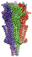
Full-length serotonin receptor (ion channel)
6579
A 3D reconstruction, created using cryo-electron microscopy, of an ion channel known as the full-length serotonin receptor in complex with the antinausea drug granisetron (orange). Sudha Chakrapani, Case Western Reserve University School of Medicine. View Media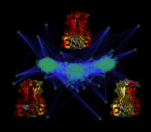
Cluster analysis of mysterious protein
3295
Researchers use cluster analysis to study protein shape and function. Each green circle represents one potential shape of the protein mitoNEET. Patricia Jennings and Elizabeth Baxter, University of California, San Diego View Media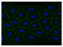
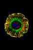
Draper, shown in the fatbody of a Drosophila melanogaster larva
2757
The fly fatbody is a nutrient storage and mobilization organ akin to the mammalian liver. The engulfment receptor Draper (green) is located at the cell surface of fatbody cells. Christina McPhee and Eric Baehrecke, University of Massachusetts Medical School View Media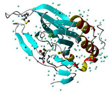
Cysteine dioxygenase from mouse
2347
Model of the mammalian iron enzyme cysteine dioxygenase from a mouse. Center for Eukaryotic Structural Genomics, PSI View Media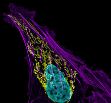
Bone cancer cell
3626
This image shows an osteosarcoma cell with DNA in blue, energy factories (mitochondria) in yellow, and actin filaments—part of the cellular skeleton—in purple. Dylan Burnette and Jennifer Lippincott-Schwartz, NICHD View Media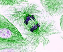
Dividing cells showing chromosomes and cell skeleton
3631
This pig cell is in the process of dividing. The chromosomes (purple) have already replicated and the duplicates are being pulled apart by fibers of the cell skeleton known as microtubules (green). Nasser Rusan, National Heart, Lung, and Blood Institute, National Institutes of Health View Media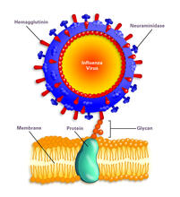
Influenza virus attaches to host membrane (with labels)
2505
Influenza A infects a host cell when hemagglutinin grips onto glycans on its surface. Crabtree + Company View Media
Leading cells with light
2708
A blue laser beam turns on a protein that helps this human cancer cell move. Responding to the stimulus, the protein, called Rac1, first creates ruffles at the edge of the cell. Yi Wu, University of North Carolina View Media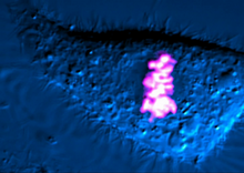
Dividing cell
6965
As this cell was undergoing cell division, it was imaged with two microscopy techniques: differential interference contrast (DIC) and confocal. The DIC view appears in blue and shows the entire cell. Dylan T. Burnette, Vanderbilt University School of Medicine. View Media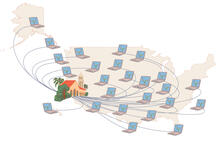
Folding@Home
1276
Stanford University scientist Vijay Pande decided to couple the power of computers with the help of the public. Judith Stoffer View Media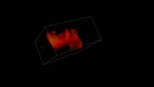
Misfolded proteins in mitochondria, 3-D video
5877
Three-dimensional image of misfolded proteins (green) within mitochondria (red). Related to image 5878. Rong Li, Department of Chemical and Biomolecular Engineering, Whiting School of Engineering, Johns Hopkins University View Media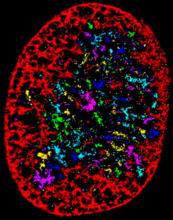
Chromatin in human fibroblast
6887
The nucleus of a human fibroblast cell with chromatin—a substance made up of DNA and proteins—shown in various colors. Melike Lakadamyali, Perelman School of Medicine at the University of Pennsylvania. View Media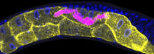
Microsporidia in roundworm 1
5777
Many disease-causing microbes manipulate their host’s metabolism and cells for their own ends. Keir Balla and Emily Troemel, University of California San Diego View Media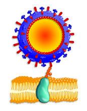
Influenza virus attaches to host membrane
2425
Influenza A infects a host cell when hemagglutinin grips onto glycans on its surface. Crabtree + Company View Media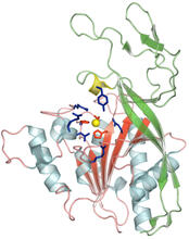
Human aspartoacylase
2352
Model of aspartoacylase, a human enzyme involved in brain metabolism. Center for Eukaryotic Structural Genomics, PSI View Media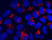
Suicidal Stem Cells
3341
Embryonic stem cells store pre-activated Bax (red) in the Golgi, near the nucleus (blue). Featured in the June 21, 2012, issue of Biomedical Beat. Mohanish Deshmukh View Media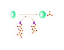
Kinases
2534
Kinases are enzymes that add phosphate groups (red-yellow structures) to proteins (green), assigning the proteins a code. Crabtree + Company View Media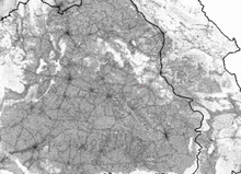
Simulation of controlled avian flu outbreak
2573
This video shows a controlled outbreak of transmissible avian flu among people living in Thailand. Neil M. Ferguson, Imperial College London View Media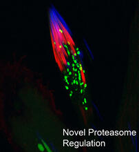
Proteasome
3451
This fruit fly spermatid recycles various molecules, including malformed or damaged proteins. Sigi Benjamin-Hong, Rockefeller University View Media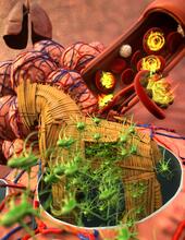
Host infection stimulates antibiotic resistance
5764
This illustration shows pathogenic bacteria behave like a Trojan horse: switching from antibiotic susceptibility to resistance during infection. View Media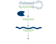
Dicer generates microRNAs
2556
The enzyme Dicer generates microRNAs by chopping larger RNA molecules into tiny Velcro®-like pieces. MicroRNAs stick to mRNA molecules and prevent the mRNAs from being made into proteins. Crabtree + Company View Media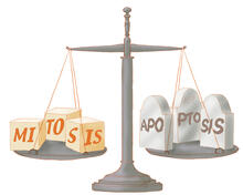
Life in balance
1336
Mitosis creates cells, and apoptosis kills them. The processes often work together to keep us healthy. Judith Stoffer View Media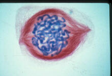
Lily mitosis 05
1015
A light microscope image of a cell from the endosperm of an African globe lily (Scadoxus katherinae). This is one frame of a time-lapse sequence that shows cell division in action. Andrew S. Bajer, University of Oregon, Eugene View Media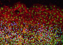
Cell proliferation in a quail embryo
2808
Image showing that the edge zone (top of image) of the quail embryo shows no proliferating cells (cyan), unlike the interior zone (bottom of image). Non-proliferating cell nuclei are labeled green. Andrés Garcia, Georgia Tech View Media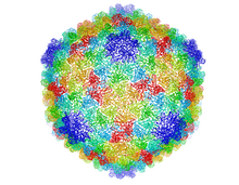
Bacteriophage P22 capsid
5874
Cryo-electron microscopy (cryo-EM) has the power to capture details of proteins and other small biological structures at the molecular level. This image shows proteins in the capsid, or outer co Dr. Wah Chiu, Baylor College of Medicine View Media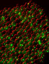
Protein clumping in zinc-deficient yeast cells
3550
The green spots in this image are clumps of protein inside yeast cells that are deficient in both zinc and a protein called Tsa1 that prevents clumping. Colin MacDiarmid and David Eide, University of Wisconsin--Madison View Media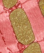
Mitochondria from rat heart muscle cell_2
3664
These mitochondria (brown) are from the heart muscle cell of a rat. Mitochondria have an inner membrane that folds in many places (and that appears here as striations). National Center for Microscopy and Imaging Research View Media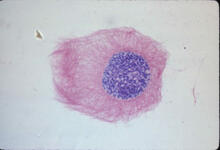
Lily mitosis 03
1013
A light microscope image of a cell from the endosperm of an African globe lily (Scadoxus katherinae). This is one frame of a time-lapse sequence that shows cell division in action. Andrew S. Bajer, University of Oregon, Eugene View Media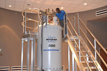
800 MHz NMR magnet
3526
Scientists use nuclear magnetic spectroscopy (NMR) to determine the detailed, 3D structures of molecules. Asokan Anbanandam, University of Kansas View Media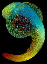
Zebrafish embryo
3644
Just 22 hours after fertilization, this zebrafish embryo is already taking shape. By 36 hours, all of the major organs will have started to form. Philipp Keller, Bill Lemon, Yinan Wan, and Kristin Branson, Janelia Farm Research Campus, Howard Hughes Medical Institute, Ashburn, Va. View Media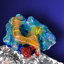
Kinesin moves cellular cargo
3491
A protein called kinesin (blue) is in charge of moving cargo around inside cells and helping them divide. Charles Sindelar, Yale University View Media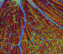
Mouse Retina
3309
A genetic disorder of the nervous system, neurofibromatosis causes tumors to form on nerves throughout the body, including a type of tumor called an optic nerve glioma that can result in childhood bli Tom Deerinck, NCMIR View Media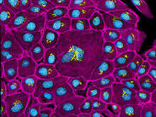
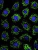
Epithelial cells
3647
This image mostly shows normal cultured epithelial cells expressing green fluorescent protein targeted to the Golgi apparatus (yellow-green) and stained for actin (magenta) and DNA (cyan). Tom Deerinck, National Center for Microscopy and Imaging Research (NCMIR) View Media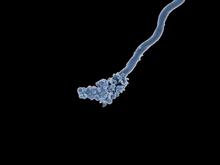
Relapsing fever bacterium (gray) and red blood cells
3585
Relapsing fever is caused by a bacterium and transmitted by certain soft-bodied ticks or body lice. The disease is seldom fatal in humans, but it can be very serious and prolonged. NIAID View Media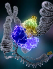
Repairing DNA
2330
Like a watch wrapped around a wrist, a special enzyme encircles the double helix to repair a broken strand of DNA. Tom Ellenberger, Washington University School of Medicine View Media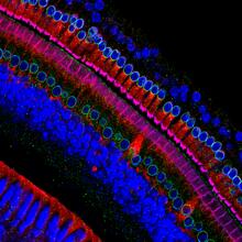
Hair cells: the sound-sensing cells in the ear
3618
These cells get their name from the hairlike structures that extend from them into the fluid-filled tube of the inner ear. Henning Horn, Brian Burke, and Colin Stewart, Institute of Medical Biology, Agency for Science, Technology, and Research, Singapore View Media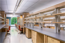
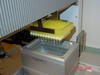
iPS cell facility at the Coriell Institute for Medical Research
2723
This lab space was designed for work on the induced pluripotent stem (iPS) cell collection, part of the NIGMS Human Genetic Cell Repository at the Coriell Institute for Medical Research. Courtney Sill, Coriell Institute for Medical Research View Media
Proteins related to myotonic dystrophy
2727
Myotonic dystrophy is thought to be caused by the binding of a protein called Mbnl1 to abnormal RNA repeats. Manuel Ares, University of California, Santa Cruz View Media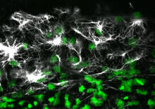
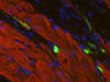
Vimentin in a quail embryo
2807
Confocal image showing high levels of the protein vimentin (white) at the edge zone of a quail embryo. Cell nuclei are labeled green. Andrés Garcia, Georgia Tech View Media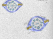
Mouse sperm sections
1191
This transmission electron micrograph shows sections of mouse sperm tails, or flagella. Tina Weatherby Carvalho, University of Hawaii at Manoa View Media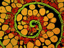
Anglerfish ovary cross-section
3620
This image captures the spiral-shaped ovary of an anglerfish in cross-section. Once matured, these eggs will be released in a gelatinous, floating mass. James E. Hayden, The Wistar Institute, Philadelphia, Pa. View Media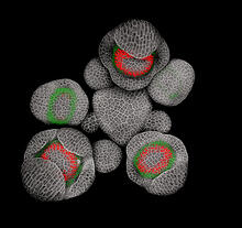
Developing Arabidopsis flower buds
3743
Flower development is a carefully orchestrated, genetically programmed process that ensures that the male (stamen) and female (pistil) organs form in the right place and at the right time in the flowe Nathanaël Prunet, Caltech View Media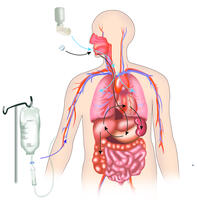
A drug's life in the body
2527
A drug's life in the body. Medicines taken by mouth pass through the liver before they are absorbed into the bloodstream. Crabtree + Company View Media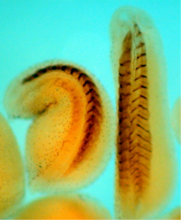
Xenopus laevis embryos
2756
Xenopus laevis, the African clawed frog, has long been used as a model organism for studying embryonic development. The frog embryo on the left lacks the developmental factor Sizzled. Michael Klymkowsky, University of Colorado, Boulder View Media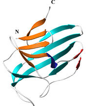
Secreted protein from Mycobacteria
2379
Model of a major secreted protein of unknown function, which is only found in mycobacteria, the class of bacteria that causes tuberculosis. Mycobacterium Tuberculosis Center, PSI View Media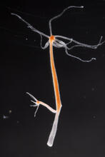
Hydra 02
2438
Hydra magnipapillata is an invertebrate animal used as a model organism to study developmental questions, for example the formation of the body axis. Hiroshi Shimizu, National Institute of Genetics in Mishima, Japan View Media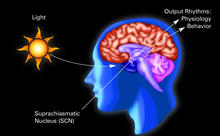
Circadian rhythm (with labels)
2569
The human body keeps time with a master clock called the suprachiasmatic nucleus or SCN. Crabtree + Company View Media