Switch to Gallery View
Image and Video Gallery
This is a searchable collection of scientific photos, illustrations, and videos. The images and videos in this gallery are licensed under Creative Commons Attribution Non-Commercial ShareAlike 3.0. This license lets you remix, tweak, and build upon this work non-commercially, as long as you credit and license your new creations under identical terms.
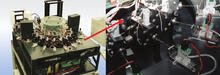
Capillary protein crystallization robot
2357
This ACAPELLA robot for capillary protein crystallization grows protein crystals, freezes them, and centers them without manual intervention. Structural Genomics of Pathogenic Protozoa Consortium View Media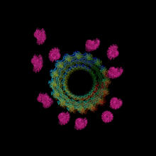
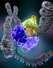
Actin filaments bundled around the dynamin helical polymer
6571
Multiple actin filaments (magenta) are organized around a dynamin helical polymer (rainbow colored) in this model derived from cryo-electron tomography. Elizabeth Chen, University of Texas Southwestern Medical Center. View Media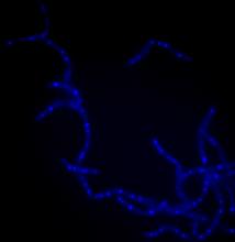
Bacillus anthracis being killed
3525
Bacillus anthracis (anthrax) cells being killed by a fluorescent trans-translation inhibitor, which disrupts bacterial protein synthesis. Kenneth Keiler, Penn State University View Media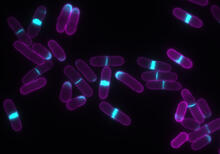
Yeast cells with accumulated cell wall material
6797
Yeast cells that abnormally accumulate cell wall material (blue) at their ends and, when preparing to divide, in their middles. This image was captured using wide-field microscopy with deconvolution. Alaina Willet, Kathy Gould’s lab, Vanderbilt University. View Media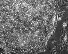
Induced stem cells from adult skin 01
2603
These cells are induced stem cells made from human adult skin cells that were genetically reprogrammed to mimic embryonic stem cells. James Thomson, University of Wisconsin-Madison View Media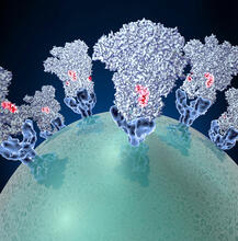
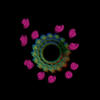
Coronavirus spike protein structure
3753
Coronaviruses are enveloped viruses responsible for 30 percent of mild respiratory infections and atypical deadly pneumonia in humans worldwide. Melody Campbell, UCSF View Media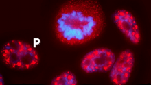
Pathways: What's the Connection? | Different Jobs in a Science Lab
6541
Learn about some of the different jobs in a scientific laboratory and how researchers work as a team to make discoveries. National Institute of General Medical Sciences View Media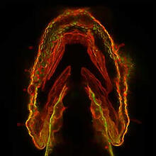
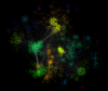
Glowing glycans
2473
Sugars light up the cells in this jaw of a 3-day-old zebrafish embryo and highlight a scientific first: labeling and tracking the movements of sugar chains called glycans in a living organism. Carolyn Bertozzi, University of California, Berkeley View Media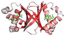
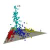
Dimeric ferredoxin-like protein from an unidentified marine microbe
2340
This is the first structure of a protein derived from the metagenomic sequences collected during the Sorcerer II Global Ocean Sampling project. Joint Center for Structural Genomics View Media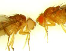
Drosophila
6344
Two adult fruit flies (Drosophila) Dr. Vicki Losick, MDI Biological Laboratory, www.mdibl.org View Media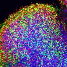
Human retinal organoid
6748
A replica of a human retina grown from stem cells. Kevin Eliceiri, University of Wisconsin-Madison. View Media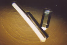
Bovine milk alpha-lactalbumin (2)
2404
Crystals of bovine milk alpha-lactalbumin protein created for X-ray crystallography, which can reveal detailed, three-dimensional protein structures. Alex McPherson, University of California, Irvine View Media
Epigenetic code
2562
The "epigenetic code" controls gene activity with chemical tags that mark DNA (purple diamonds) and the "tails" of histone proteins (purple triangles). Crabtree + Company View Media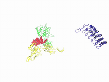
A molecular switch strips transcription factor from DNA
3729
In this video, Rice University scientists used molecular modeling with a mathematical algorithm called AWSEM (for associative memory, water-mediated, structure and energy model) and structural data to Davit Potoyan and Peter Wolynes View Media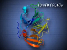
See how immune cell acid destroys bacterial proteins
6602
This animation shows the effect of exposure to hypochlorous acid, which is found in certain types of immune cells, on bacterial proteins. American Chemistry Council View Media
Lorsch Swearing In
3530
Jon Lorsch at his swearing in as NIGMS director in August 2013. Also shown are Francis Collins, NIH Director, and Judith Greenberg, former NIGMS Acting Director. View Media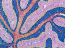
Mouse cerebellum in pink and blue
5800
The cerebellum is the brain's locomotion control center. Found at the base of your brain, the cerebellum is a single layer of tissue with deep folds like an accordion. National Center for Microscopy and Imaging Research (NCMIR) View Media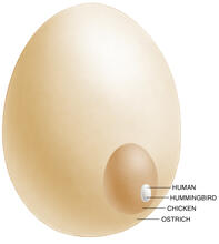
Egg comparison
1339
The largest human cell (by volume) is the egg. Human eggs are 150 micrometers in diameter and you can just barely see one with a naked eye. In comparison, consider the eggs of chickens...or ostriches! Judith Stoffer View Media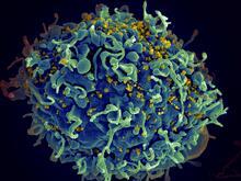
HIV, the AIDS virus, infecting a human cell
3638
This human T cell (blue) is under attack by HIV (yellow), the virus that causes AIDS. Seth Pincus, Elizabeth Fischer, and Austin Athman, National Institute of Allergy and Infectious Diseases, National Institutes of Health View Media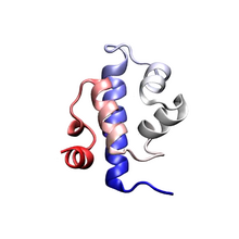
Protein folding video
3391
Proteins are long chains of amino acids. Each protein has a unique amino acid sequence. It is still a mystery how a protein folds into the proper shape based on its sequence. Theoretical and Computational Biophysics Group View Media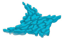
Smooth ER
1292
The endoplasmic reticulum comes in two types: Rough ER is covered with ribosomes and prepares newly made proteins; smooth ER specializes in making lipids and breaking down toxic molecules. Judith Stoffer View Media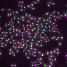
Yeast cells with nuclei and contractile rings
6792
Yeast cells with nuclei shown in green and contractile rings shown in magenta. Nuclei store DNA, and contractile rings help cells divide. Alaina Willet, Kathy Gould’s lab, Vanderbilt University. View Media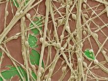
Synapses in culture
3399
Cultured hippocampal neurons grown on a substrate of glial cells (astrocytes). The glial cells form the pink/brown underlayment in this image. The tan threads are the neurons. National Center for Microscopy and Imaging Research View Media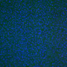
Movie of in vitro assembly of a cell-signaling pathway
3786
T cells are white blood cells that are important in defending the body against bacteria, viruses and other pathogens. Xiaolei Su, HHMI Whitman Center of the Marine Biological Laboratory View Media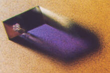
Rabbit GPDA
2405
A crystal of rabbit GPDA protein created for X-ray crystallography, which can reveal detailed, three-dimensional protein structures. Alex McPherson, University of California, Irvine View Media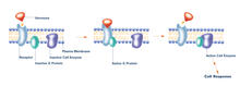
G switch (with labels)
2537
The G switch allows our bodies to respond rapidly to hormones. G proteins act like relay batons to pass messages from circulating hormones into cells. Crabtree + Company View Media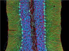
Mouse cerebellum close-up
3371
The cerebellum is the brain's locomotion control center. Every time you shoot a basketball, tie your shoe or chop an onion, your cerebellum fires into action. National Center for Microscopy and Imaging Research (NCMIR) View Media
Natcher Building 10
1090
NIGMS staff are located in the Natcher Building on the NIH campus. Alisa Machalek, National Institute of General Medical Sciences View Media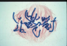
Lily mitosis 06
1016
A light microscope image of a cell from the endosperm of an African globe lily (Scadoxus katherinae). This is one frame of a time-lapse sequence that shows cell division in action. Andrew S. Bajer, University of Oregon, Eugene View Media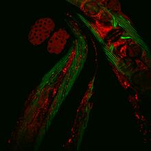
Closeup of fluorescent C. elegans showing muscle and ribosomal protein
6583
Closeup of C. elegans, tiny roundworms, with a ribosomal protein glowing red and muscle fibers glowing green. Researchers used these worms to study a molecular pathway that affects aging. Jarod Rollins, Mount Desert Island Biological Laboratory. View Media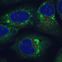
Endoplasmic reticulum abnormalities
6773
Human cells with the gene that codes for the protein FIT2 deleted. Green indicates an endoplasmic reticulum (ER) resident protein. Michel Becuwe, Harvard University. View Media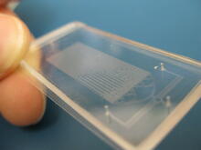
Microfluidic chip
3265
Microfluidic chips have many uses in biology labs. Jeff Hasty Lab, UC San Diego View Media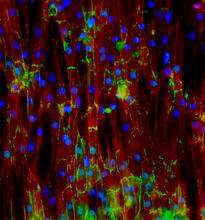
Mouse heart muscle cells
3282
This image shows neonatal mouse heart cells. These cells were grown in the lab on a chip that aligns the cells in a way that mimics what is normally seen in the body. Kara McCloskey lab, University of California, Merced, via CIRM View Media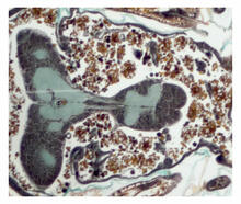
Cross section of a Drosophila melanogaster pupa
2758
This photograph shows a magnified view of a Drosophila melanogaster pupa in cross section. Compare this normal pupa to one that lacks an important receptor, shown in image 2759. Christina McPhee and Eric Baehrecke, University of Massachusetts Medical School View Media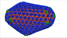
HIV Capsid
3477
This image is a computer-generated model of the approximately 4.2 million atoms of the HIV capsid, the shell that contains the virus' genetic material. Juan R. Perilla and the Theoretical and Computational Biophysics Group, University of Illinois at Urbana-Champaign View Media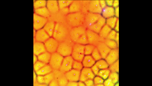
Cell-like compartments emerging from scrambled frog eggs
6587
Cell-like compartments spontaneously emerge from scrambled frog eggs, with nuclei (blue) from frog sperm. Endoplasmic reticulum (red) and microtubules (green) are also visible. Xianrui Cheng, Stanford University School of Medicine. View Media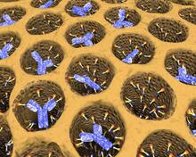
Antibodies in silica honeycomb
2750
Antibodies are among the most promising therapies for certain forms of cancer, but patients must take them intravenously, exposing healthy tissues to the drug and increasing the risk of side effects. Chenghong Lei, Pacific Northwest National Laboratory & Karl Erik Hellstrom, University of Washington View Media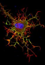
Abnormal, spiky fibroblast
3613
This is a fibroblast, a connective tissue cell that plays an important role in wound healing. Normal fibroblasts have smooth edges. Praveen Suraneni, Stowers Institute for Medical Research, Kansas City, Mo. View Media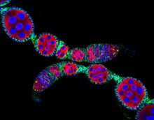
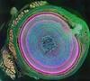
Confocal microscopy image of two Drosophila ovarioles
5772
Ovarioles in female insects are tubes in which egg cells (called oocytes) form at one end and complete their development as they reach the other end of the tube. 2004 Olympus BioScapes Competition View Media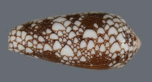
Cone snail shell
2576
A shell from the venomous cone snail Conus omaria, which lives in the Pacific and Indian oceans and eats other snails. Kerry Matz, University of Utah View Media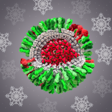
H1N1 Influenza Virus
6355
CellPack image of the H1N1 influenza virus, with hemagglutinin and neuraminidase glycoproteins in green and red, respectively, on the outer envelope (white); matrix protein in gray, and ribonucleoprot Dr. Rommie Amaro, University of California, San Diego View Media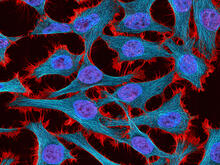
HeLa cells
3521
Multiphoton fluorescence image of HeLa cells stained with the actin binding toxin phalloidin (red), microtubules (cyan) and cell nuclei (blue). Nikon RTS2000MP custom laser scanning microscope. National Center for Microscopy and Imaging Research (NCMIR) View Media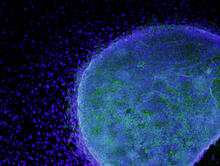
Colony of human ES cells
3269
A colony of human embryonic stem cells (light blue) grows on fibroblasts (dark blue). California Institute for Regenerative Medicine View MediaBeta-galactosidase montage showing cryo-EM improvement--transparent background
5882
Composite image of beta-galactosidase showing how cryo-EM’s resolution has improved dramatically in recent years. Older images to the left, more recent to the right. Veronica Falconieri, Sriram Subramaniam Lab, National Cancer Institute View Media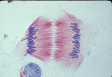
Lily mitosis 11
1011
A light microscope image of cells from the endosperm of an African globe lily (Scadoxus katherinae). This is one frame of a time-lapse sequence that shows cell division in action. Andrew S. Bajer, University of Oregon, Eugene View Media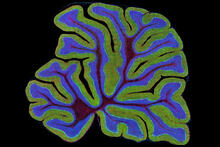
Cerebellum: the brain's locomotion control center
3639
The cerebellum of a mouse is shown here in cross-section. The cerebellum is the brain's locomotion control center. Thomas Deerinck, National Center for Microscopy and Imaging Research, University of California, San Diego View Media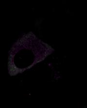
Stress Response in Cells
6570
Two highly stressed osteosarcoma cells are shown with a set of green droplet-like structures followed by a second set of magenta droplets. Julia F. Riley and Carlos A. Castañeda, Syracuse University View Media
Aging book of life
1334
Damage to each person's genome, often called the "Book of Life," accumulates with time. Judith Stoffer View Media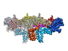
Dengue virus membrane protein structure
3758
Dengue virus is a mosquito-borne illness that infects millions of people in the tropics and subtropics each year. Like many viruses, dengue is enclosed by a protective membrane. Hong Zhou, UCLA View Media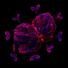
Wild-type and mutant fruit fly ovaries
6806
The two large, central, round shapes are ovaries from a typical fruit fly (Drosophila melanogaster). Vladimir I. Gelfand, Feinberg School of Medicine, Northwestern University. View Media