Switch to Gallery View
Image and Video Gallery
This is a searchable collection of scientific photos, illustrations, and videos. The images and videos in this gallery are licensed under Creative Commons Attribution Non-Commercial ShareAlike 3.0. This license lets you remix, tweak, and build upon this work non-commercially, as long as you credit and license your new creations under identical terms.
CRISPR
6351
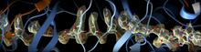
RNA incorporated into the CRISPR surveillance complex is positioned to scan across foreign DNA. Cryo-EM density from a 3Å reconstruction is shown as a yellow mesh. NRAMM National Resource for Automated Molecular Microscopy http://nramm.nysbc.org/nramm-images/ Source: Bridget Carragher View Media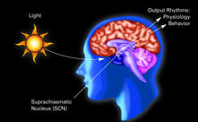
Circadian rhythm (with labels)
2569
The human body keeps time with a master clock called the suprachiasmatic nucleus or SCN. Crabtree + Company View Media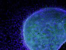
Colony of human ES cells
3269
A colony of human embryonic stem cells (light blue) grows on fibroblasts (dark blue). California Institute for Regenerative Medicine View Media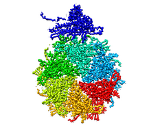
Bacteriophage P22 capsid, detail
5875
Detail of a subunit of the capsid, or outer cover, of bacteriophage P22, a virus that infects the Salmonella bacteria. Dr. Wah Chiu, Baylor College of Medicine View Media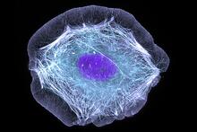
Skin cell (keratinocyte)
3599
This normal human skin cell was treated with a growth factor that triggered the formation of specialized protein structures that enable the cell to move. Torsten Wittmann, University of California, San Francisco View Media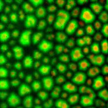
Cell-like compartments from frog eggs 3
6586
Cell-like compartments that spontaneously emerged from scrambled frog eggs. Endoplasmic reticulum (red) and microtubules (green) are visible. Image created using epifluorescence microscopy. Xianrui Cheng, Stanford University School of Medicine. View Media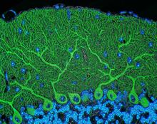
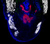
Mouse cerebellum
5795
The cerebellum is the brain's locomotion control center. Found at the base of your brain, the cerebellum is a single layer of tissue with deep folds like an accordion. National Center for Microscopy and Imaging Research (NCMIR) View Media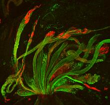
Fruit fly spermatids
3590
Developing spermatids (precursors of mature sperm cells) begin as small, round cells and mature into long-tailed, tadpole-shaped ones. Lacramioara Fabian, The Hospital for Sick Children, Toronto, Canada View Media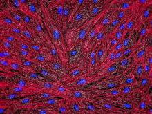
DNA and actin in cultured fibroblast cells
3670
DNA (blue) and actin (red) in cultured fibroblast cells. Tom Deerinck, National Center for Microscopy and Imaging Research (NCMIR) View Media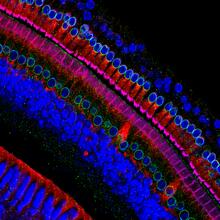
Hair cells: the sound-sensing cells in the ear
3618
These cells get their name from the hairlike structures that extend from them into the fluid-filled tube of the inner ear. Henning Horn, Brian Burke, and Colin Stewart, Institute of Medical Biology, Agency for Science, Technology, and Research, Singapore View Media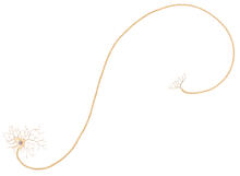
Nerve cell
1338
Nerve cells have long, invisibly thin fibers that carry electrical impulses throughout the body. Some of these fibers extend about 3 feet from the spinal cord to the toes. Judith Stoffer View Media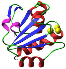
PanB from M. tuberculosis (2)
2382
Model of an enzyme, PanB, from Mycobacterium tuberculosis, the bacterium that causes most cases of tuberculosis. This enzyme is an attractive drug target. Mycobacterium Tuberculosis Center, PSI-1 View Media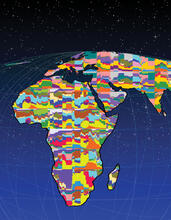
Mapping human genetic variation
2443
This map paints a colorful portrait of human genetic variation around the world. Noah Rosenberg and Martin Soave, University of Michigan View Media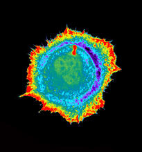
Seeing signaling protein activation in cells 01
2451
Cdc42, a member of the Rho family of small guanosine triphosphatase (GTPase) proteins, regulates multiple cell functions, including motility, proliferation, apoptosis, and cell morphology. Klaus Hahn, University of North Carolina, Chapel Hill Medical School View Media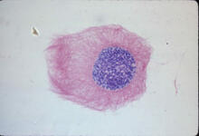
Lily mitosis 03
1013
A light microscope image of a cell from the endosperm of an African globe lily (Scadoxus katherinae). This is one frame of a time-lapse sequence that shows cell division in action. Andrew S. Bajer, University of Oregon, Eugene View Media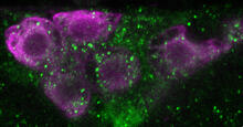
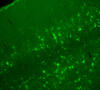
Insulin production and fat sensing in fruit flies
6982
Fourteen neurons (magenta) in the adult Drosophila brain produce insulin, and fat tissue sends packets of lipids to the brain via the lipoprotein carriers (green). Akhila Rajan, Fred Hutchinson Cancer Center View Media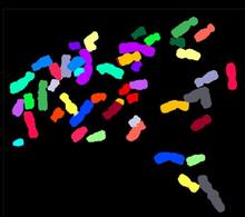
Color-coded chromosomes
2312
By mixing fluorescent dyes like an artist mixes paints, scientists are able to color code individual chromosomes. Anna Jauch, Institute of Human Genetics, Heidelberg, Germany View Media
Katanin protein regulates anaphase
2594
The microtubule severing protein, katanin, localizes to chromosomes and regulates anaphase A in mitosis. David Sharp, Albert Einstein College of Medicine View Media
Planting roots
2329
At the root tips of the mustard plant Arabidopsis thaliana (red), two proteins work together to control the uptake of water and nutrients. Philip Benfey, Duke University View Media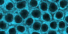
Movements of myosin
2324
Inside the fertilized egg cell of a fruit fly, we see a type of myosin (related to the protein that helps muscles contract) made to glow by attaching a fluorescent protein. Victoria Foe, University of Washington View Media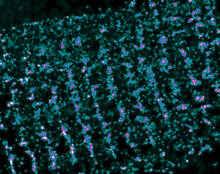
Single-Molecule Imaging
3339
This is a super-resolution light microscope image taken by Hiro Hakozaki and Masa Hoshijima of NCMIR. Tom Deerinck, NCMIR View Media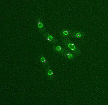
Dividing yeast cells with nuclear envelopes and spindle pole bodies
6795
Time-lapse video of yeast cells undergoing cell division. Nuclear envelopes are shown in green, and spindle pole bodies, which help pull apart copied genetic information, are shown in magenta. Alaina Willet, Kathy Gould’s lab, Vanderbilt University. View Media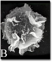
Activated mast cell surface
2637
A scanning electron microscope image of an activated mast cell. This image illustrates the interesting topography of the cell membrane, which is populated with receptors. Bridget Wilson, University of New Mexico View Media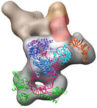
Structure of telomerase
3459
Scientists recently discovered the full molecular structure of telomerase, an enzyme important to aging and cancer. Jiansen Jiang, Edward J. Miracco, Z. Hong Zhou and Juli Feigon, University of California, Los Angeles; Kathleen Collins, University of California, Berkeley View Media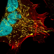
Multinucleated cancer cell
6967
A cancer cell with three nuclei, shown in turquoise. The abnormal number of nuclei indicates that the cell failed to go through cell division, probably more than once. Dylan T. Burnette, Vanderbilt University School of Medicine. View Media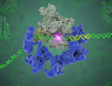
TFIID complex binds DNA to start gene transcription
3766
Gene transcription is a process by which the genetic information encoded in DNA is transcribed into RNA. Eva Nogales, Berkeley Lab View Media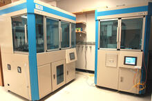
Cell-free protein synthesizers
2360
Both instruments shown were developed by CellFree Sciences of Yokohama, Japan. Center for Eukaryotic Structural Genomics View Media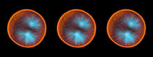
Sea urchin embryo 03
1049
Stereo triplet of a sea urchin embryo stained to reveal actin filaments (orange) and microtubules (blue). George von Dassow, University of Washington View Media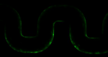
Biofilm blocking fluid flow
3446
This time-lapse movie shows that bacterial communities called biofilms can create blockages that prevent fluid flow in devices such as stents and catheters over a period of about 56 hours. Bonnie Bassler, Princeton University View Media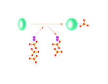
Kinases
2534
Kinases are enzymes that add phosphate groups (red-yellow structures) to proteins (green), assigning the proteins a code. Crabtree + Company View Media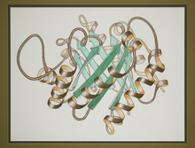
Early ribbon drawing of a protein
2748
This ribbon drawing of a protein hand drawn and colored by researcher Jane Richardson in 1981 helped originate the ribbon representation of proteins that is now ubiquitous in molecular graphics. Jane Richardson, Duke University Medical Center View Media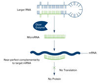
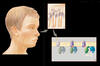
Dicer generates microRNAs (with labels)
2557
The enzyme Dicer generates microRNAs by chopping larger RNA molecules into tiny Velcro®-like pieces. MicroRNAs stick to mRNA molecules and prevent the mRNAs from being made into proteins. Crabtree + Company View Media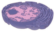
Nucleus and rough ER
1290
The nucleus contains the DNA of eukaryotic cells. Judith Stoffer View Media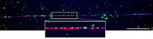
Chromosome fiber 01
2475
This microscopic image shows a chromatin fiber--a DNA molecule bound to naturally occurring proteins. Marc Green and Susan Forsburg, University of Southern California View Media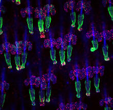
Wound healing in process
3498
Wound healing requires the action of stem cells. Hermann Steller, Rockefeller University View Media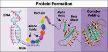
Protein formation
6603
Proteins are 3D structures made up of smaller units. DNA is transcribed to RNA, which in turn is translated into amino acids. NIGMS, with the folded protein illustration adapted from Jane Richardson, Duke University Medical Center View Media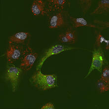
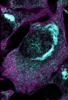
Endoplasmic reticulum abnormalities 2
6774
Human cells with the gene that codes for the protein FIT2 deleted. After an experimental intervention, they are expressing a nonfunctional version of FIT2, shown in green. Michel Becuwe, Harvard University. View Media
An adult Hawaiian bobtail squid
7013
An adult female Hawaiian bobtail squid, Euprymna scolopes, with its mantle cavity exposed from the underside. Margaret J. McFall-Ngai, Carnegie Institution for Science/California Institute of Technology, and Edward G. Ruby, California Institute of Technology. View Media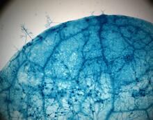
Disease-susceptible Arabidopsis leaf
2782
This is a magnified view of an Arabidopsis thaliana leaf after several days of infection with the pathogen Hyaloperonospora arabidopsidis. Jeff Dangl, University of North Carolina, Chapel Hill View Media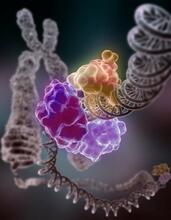
Repairing DNA
3493
Like a watch wrapped around a wrist, a special enzyme encircles the double helix to repair a broken strand of DNA. Tom Ellenberger, Washington University School of Medicine View Media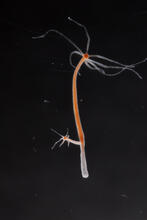
Hydra 04
2440
Hydra magnipapillata is an invertebrate animal used as a model organism to study developmental questions, for example the formation of the body axis. Hiroshi Shimizu, National Institute of Genetics in Mishima, Japan View Media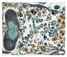
Cross section of a Drosophila melanogaster pupa lacking Draper
2759
In the absence of the engulfment receptor Draper, salivary gland cells (light blue) persist in the thorax of a developing Drosophila melanogaster pupa. Christina McPhee and Eric Baehrecke, University of Massachusetts Medical School View Media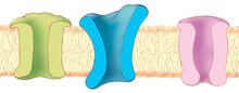
Ion channels
1284
The body uses a variety of ion channels to transport small molecules across cell membranes. Judith Stoffer View Media
Network Map
2735
This network map shows the overlap (green) between the long QT syndrome (yellow) and epilepsy (blue) protein-interaction neighborhoods located within the human interactome. Seth Berger, Mount Sinai School of Medicine View Media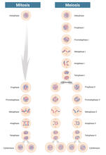
Mitosis and meiosis compared-labeled
6788
Meiosis is used to make sperm and egg cells. During meiosis, a cell's chromosomes are copied once, but the cell divides twice. Judith Stoffer View Media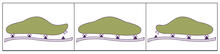
Focal adhesions
2502
Cells walk along body surfaces via tiny "feet," called focal adhesions, that connect with the extracellular matrix. Crabtree + Company View Media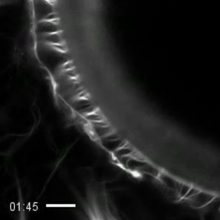
How cilia do the wave
3494
Thin, hair-like biological structures called cilia are tiny but mighty. Zvonimir Dogic, Brandeis University View Media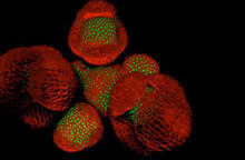
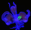
Arabidopsis Thaliana: Flowers Spring to Life
6503
This image capture shows how a single gene, STM, plays a starring role in plant development. Nathanaёl Prunet NIH Support: National Institute of General Medical Sciences View Media
Stretch detectors
2714
Muscles stretch and contract when we walk, and skin splits open and knits back together when we get a paper cut. Christopher Chen, University of Pennsylvania View Media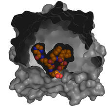
X-ray co-crystal structure of Src kinase bound to a DNA-templated macrocycle inhibitor 5
3417
X-ray co-crystal structure of Src kinase bound to a DNA-templated macrocycle inhibitor. Markus A. Seeliger, Stony Brook University Medical School and David R. Liu, Harvard University View Media