
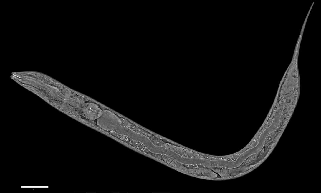
Image Gallery: C. elegans showing internal structures
ID
6961
An image of Caenorhabditis elegans, a tiny roundworm, showing internal structures including the intestine, pharynx, and body wall muscle. C. elegans is one of the simplest organisms with a nervous system. Scientists use it to study nervous system development, among other things. This image was captured with a quantitative orientation-independent differential interference contrast (OI-DIC) microscope. The scale bar is 100 µm.
More information about the microscopy that produced this image can be found in the Journal of Microscopy paper by Malamy and Shribak.
More information about the microscopy that produced this image can be found in the Journal of Microscopy paper by Malamy and Shribak.
Source
Michael Shribak, Marine Biological Laboratory/University of Chicago.
Topics

