Switch to Gallery View
Image and Video Gallery
This is a searchable collection of scientific photos, illustrations, and videos. The images and videos in this gallery are licensed under Creative Commons Attribution Non-Commercial ShareAlike 3.0. This license lets you remix, tweak, and build upon this work non-commercially, as long as you credit and license your new creations under identical terms.
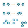
Fruit fly egg ooplasmic streaming
6809
Two fruit fly (Drosophila melanogaster) egg cells, one on each side of the central black line. Vladimir I. Gelfand, Feinberg School of Medicine, Northwestern University. View Media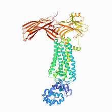
Rhodopsin bound to visual arrestin
6768
Rhodopsin is a pigment in the rod cells of the retina (back of the eye). It is extremely light-sensitive, supporting vision in low-light conditions. Protein Data Bank. View Media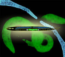
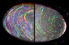
Worms and human infertility
2333
This montage of tiny, transparent C. elegans--or roundworms--may offer insight into understanding human infertility. Abby Dernburg, Lawrence Berkeley National Laboratory View Media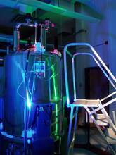
Superconducting magnet
1120
Superconducting magnet for NMR research, from the February 2003 profile of Dorothee Kern in Findings. Mike Lovett View Media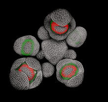
Developing Arabidopsis flower buds
3743
Flower development is a carefully orchestrated, genetically programmed process that ensures that the male (stamen) and female (pistil) organs form in the right place and at the right time in the flowe Nathanaël Prunet, Caltech View Media
Natcher Building 10
1090
NIGMS staff are located in the Natcher Building on the NIH campus. Alisa Machalek, National Institute of General Medical Sciences View Media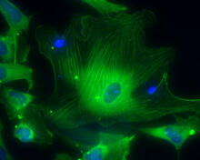
Smooth muscle from human ES cells
3288
These smooth muscle cells were derived from human embryonic stem cells. The nuclei are stained blue, and the proteins of the cytoskeleton are stained green. Alexey Terskikh lab, Burnham Institute for Medical Research, via CIRM View Media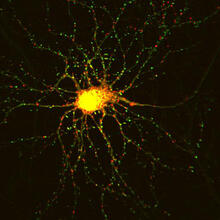
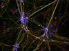
Neuron with labeled synapses
3509
In this image, recombinant probes known as FingRs (Fibronectin Intrabodies Generated by mRNA display) were expressed in a cortical neuron, where they attached fluorescent proteins to either PSD95 (gre Don Arnold and Richard Roberts, University of Southern California. View Media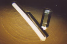
Bovine milk alpha-lactalbumin (2)
2404
Crystals of bovine milk alpha-lactalbumin protein created for X-ray crystallography, which can reveal detailed, three-dimensional protein structures. Alex McPherson, University of California, Irvine View Media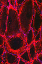
Cells lining the blood vessel walls
3633
The structure of the endothelium, the thin layer of cells that line our arteries and veins, is visible here. Christopher V. Carman and Roberta Martinelli, Harvard Medical School. View Media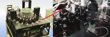
Capillary protein crystallization robot
2357
This ACAPELLA robot for capillary protein crystallization grows protein crystals, freezes them, and centers them without manual intervention. Structural Genomics of Pathogenic Protozoa Consortium View Media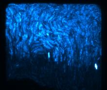
Fluorescent E. coli bacteria
3268
Bioengineers were able to coax bacteria to blink in unison on microfluidic chips. They called each blinking bacterial colony a biopixel. Thousands of fluorescent E. Jeff Hasty Lab, UC San Diego View Media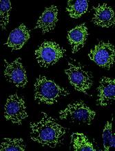
Calcium uptake during ATP production in mitochondria
3449
Living primary mouse embryonic fibroblasts. Mitochondria (green) stained with the mitochondrial membrane potential indicator, rhodamine 123. Nuclei (blue) are stained with DAPI. Lili Guo, Perelman School of Medicine, University of Pennsylvania View Media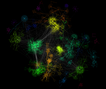
A molecular interaction network in yeast 2
3732
The image visualizes a part of the yeast molecular interaction network. Keiichiro Ono, UCSD View Media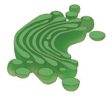
Golgi
1275
The Golgi complex, also called the Golgi apparatus or, simply, the Golgi. Judith Stoffer View Media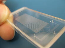
Microfluidic chip
3265
Microfluidic chips have many uses in biology labs. Jeff Hasty Lab, UC San Diego View Media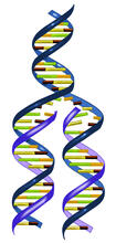
DNA replication illustration
2543
During DNA replication, each strand of the original molecule acts as a template for the synthesis of a new, complementary DNA strand. Crabtree + Company View Media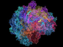
RNA Polymerase II
2484
NIGMS-funded researchers led by Roger Kornberg solved the structure of RNA polymerase II. David Bushnell, Ken Westover and Roger Kornberg, Stanford University View Media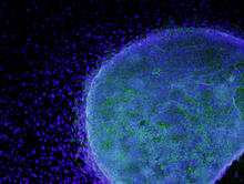
Colony of human ES cells
3269
A colony of human embryonic stem cells (light blue) grows on fibroblasts (dark blue). California Institute for Regenerative Medicine View Media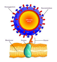
Influenza virus attaches to host membrane (with labels)
2505
Influenza A infects a host cell when hemagglutinin grips onto glycans on its surface. Crabtree + Company View Media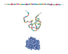
Building blocks and folding of proteins
2508
Proteins are made of amino acids hooked end-to-end like beads on a necklace. To become active, proteins must twist and fold into their final, or "native," conformation. Crabtree + Company View Media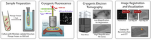
Correlative imaging by annotation with single molecules (CIASM) process
6568
These images illustrate a technique combining cryo-electron tomography and super-resolution fluorescence microscopy called correlative imaging by annotation with single molecules (CIASM). Peter Dahlberg, Stanford University. View Media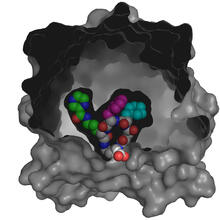
X-ray co-crystal structure of Src kinase bound to a DNA-templated macrocycle inhibitor 4
3416
X-ray co-crystal structure of Src kinase bound to a DNA-templated macrocycle inhibitor. Markus A. Seeliger, Stony Brook University Medical School and David R. Liu, Harvard University View Media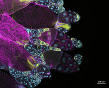
Fruit fly ovary
6522
In this image of a stained fruit fly ovary, the ovary is packed with immature eggs (with DNA stained blue). The cytoskeleton (in pink) is a collection of fibers that gives a cell shape and support. Crystal D. Rogers, Ph.D., University of California, Davis, School of Veterinary Medicine; and Mariano A. Loza-Coll, Ph.D., California State University, Northridge. View Media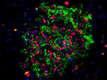
Induced pluripotent stem cells from skin
3278
These induced pluripotent stem cells (iPS cells) were derived from a woman's skin. Green and red indicate proteins found in reprogrammed cells but not in skin cells (TRA1-62 and NANOG). Kathrin Plath lab, University of California, Los Angeles, via CIRM View Media
Precisely Delivering Chemical Cargo to Cells
3779
Moving protein or other molecules to specific cells to treat or examine them has been a major biological challenge. Nature Nanotechnology View Media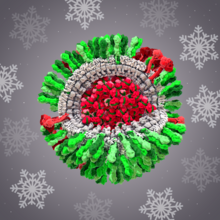
H1N1 Influenza Virus
6355
CellPack image of the H1N1 influenza virus, with hemagglutinin and neuraminidase glycoproteins in green and red, respectively, on the outer envelope (white); matrix protein in gray, and ribonucleoprot Dr. Rommie Amaro, University of California, San Diego View Media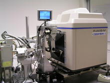
Chromium X-ray source
2361
In the determination of protein structures by X-ray crystallography, this unique soft (l = 2.29Å) X-ray source is used to collect anomalous scattering data from protein crystals containing light atoms The Southeast Collaboratory for Structural Genomics View Media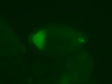
Fly cells live
2315
If a picture is worth a thousand words, what's a movie worth? Denise Montell, Johns Hopkins University School of Medicine View Media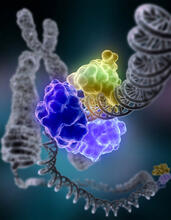
Repairing DNA
2330
Like a watch wrapped around a wrist, a special enzyme encircles the double helix to repair a broken strand of DNA. Tom Ellenberger, Washington University School of Medicine View Media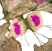
Human fibroblast undergoing cell division
6519
During cell division, cells physically divide after separating their genetic material to create two daughter cells that are genetically identical to the parent cell. Nilay Taneja, Vanderbilt University, and Dylan T. Burnette, Ph.D., Vanderbilt University School of Medicine. View Media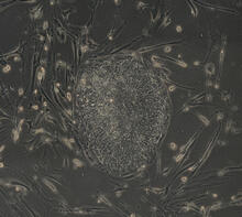
Induced stem cells from adult skin 04
2606
The human skin cells pictured contain genetic modifications that make them pluripotent, essentially equivalent to embryonic stem cells. James Thomson, University of Wisconsin-Madison View Media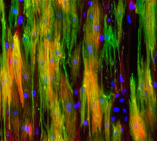
Mouse heart muscle cells 02
3283
This image shows neonatal mouse heart cells. These cells were grown in the lab on a chip that aligns the cells in a way that mimics what is normally seen in the body. Kara McCloskey lab, University of California, Merced, via CIRM View Media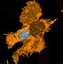
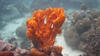
Zebrafish pigment cell
5754
Pigment cells are cells that give skin its color. David Parichy, University of Washington View Media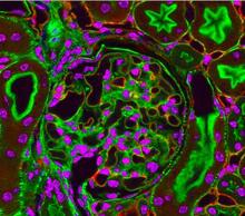
Fluorescent microscopy of kidney tissue--close-up
3725
This photograph of kidney tissue, taken using fluorescent light microscopy, shows a close-up view of part of image 3723. Tom Deerinck , National Center for Microscopy and Imaging Research View Media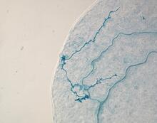
Arabidopsis leaf injected with a pathogen
2780
This is a magnified view of an Arabidopsis thaliana leaf eight days after being infected with the pathogen Hyaloperonospora arabidopsidis, which is closely related to crop pathogens that Jeff Dangl, University of North Carolina, Chapel Hill View Media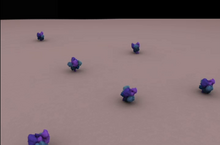
Clathrin-mediated endocytosis
5753
Endocytosis is the process by which cells are able to take up membrane and extracellular materials through the formation of a small intracellular bubble, called a vesicle. Janet Iwasa, University of Utah View Media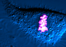
Dividing cell
6965
As this cell was undergoing cell division, it was imaged with two microscopy techniques: differential interference contrast (DIC) and confocal. The DIC view appears in blue and shows the entire cell. Dylan T. Burnette, Vanderbilt University School of Medicine. View Media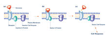
G switch (with labels and stages)
2538
The G switch allows our bodies to respond rapidly to hormones. G proteins act like relay batons to pass messages from circulating hormones into cells. Crabtree + Company View Media
Wreath-shaped protein from X. campestris
2372
Crystal structure of a protein with unknown function from Xanthomonas campestris, a plant pathogen. Eight copies of the protein crystallized to form a ring. Ken Schwinn and Sonia Espejon-Reynes, New York SGX Research Center for Structural Genomics View Media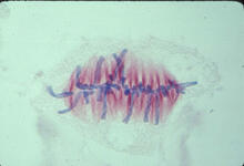
Lily mitosis 07
1017
A light microscope image of a cell from the endosperm of an African globe lily (Scadoxus katherinae). This is one frame of a time-lapse sequence that shows cell division in action. Andrew S. Bajer, University of Oregon, Eugene View Media
Z rings in bacterial division
2456
Lab-made liposomes contract where Z rings have gathered together and the constriction forces are greatest (arrows). Masaki Osawa, Duke University View Media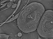
Crane fly spermatocyte undergoing meiosis
6898
A crane fly spermatocyte during metaphase of meiosis-I, a step in the production of sperm. Michael Shribak, Marine Biological Laboratory/University of Chicago. View Media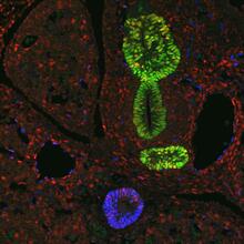
Transcription factor Sox17 controls embryonic development of certain internal organs
3440
During embryonic development, transcription factors (proteins that regulate gene expression) govern the differentiation of cells into separate tissues and organs. James M. Wells, Cincinnati Children's Hospital Medical Center View Media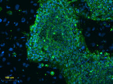
Human embryonic stem cells on feeder cells
3274
This fluorescent microscope image shows human embryonic stem cells whose nuclei are stained green. Blue staining shows the surrounding supportive feeder cells. Michael Longaker lab, Stanford University School of Medicine, via CIRM View Media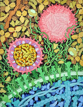
Zika virus
6998
Zika virus is shown in cross section at center left. On the outside, it includes envelope protein (red) and membrane protein (magenta) embedded in a lipid membrane (light purple). Amy Wu and Christine Zardecki, RCSB Protein Data Bank. View Media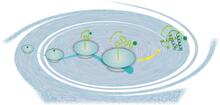
Early life of a protein
2740
This illustration represents the early life of a protein—specifically, apomyoglobin—as it is synthesized by a ribosome and emerges from the ribosomal tunnel, which contains the newly formed protein's Silvia Cavagnero, University of Wisconsin, Madison View Media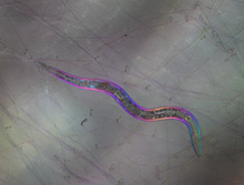
C. elegans trapped by carnivorous fungus
6963
Real-time footage of Caenorhabditis elegans, a tiny roundworm, trapped by a carnivorous fungus, Arthrobotrys dactyloides. Michael Shribak, Marine Biological Laboratory/University of Chicago. View Media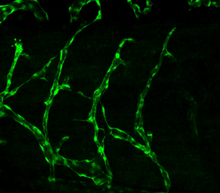
Disrupted vascular development in frog embryos
3403
Disassembly of vasculature in kdr:GFP frogs following addition of 250 µM TBZ. Related to images 3404 and 3505. Hye Ji Cha, University of Texas at Austin View Media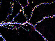
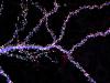
Hippocampal neuron from rodent brain
3686
Hippocampal neuron from rodent brain with dendrites shown in blue. The hundreds of tiny magenta, green and white dots are the dendritic spines of excitatory synapses. Shelley Halpain, UC San Diego View Media