Switch to Gallery View
Image and Video Gallery
This is a searchable collection of scientific photos, illustrations, and videos. The images and videos in this gallery are licensed under Creative Commons Attribution Non-Commercial ShareAlike 3.0. This license lets you remix, tweak, and build upon this work non-commercially, as long as you credit and license your new creations under identical terms.
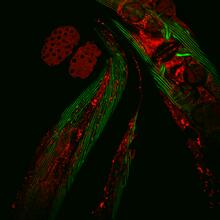
Closeup of fluorescent C. elegans showing muscle and ribosomal protein
6583
Closeup of C. elegans, tiny roundworms, with a ribosomal protein glowing red and muscle fibers glowing green. Researchers used these worms to study a molecular pathway that affects aging. Jarod Rollins, Mount Desert Island Biological Laboratory. View Media
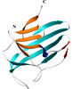
Map of protein structures 02
2367
A global "map of the protein structure universe" indicating the positions of specific proteins. Berkeley Structural Genomics Center, PSI View Media
Focal adhesions
2502
Cells walk along body surfaces via tiny "feet," called focal adhesions, that connect with the extracellular matrix. Crabtree + Company View Media
Cells lining the trachea
3646
In this image, viewed with a ZEISS ORION NanoFab microscope, the community of cells lining a mouse airway is magnified more than 10,000 times. Eva Mutunga and Kate Klein, University of the District of Columbia and National Institute of Standards and Technology View Media
Magnesium transporter protein from E. faecalis
2345
Structure of a magnesium transporter protein from an antibiotic-resistant bacterium (Enterococcus faecalis) found in the human gut. New York Structural GenomiX Consortium View MediaArtificial cilia exhibit spontaneous beating
3344
Researchers have created artificial cilia that wave like the real thing. Zvonimir Dogic View Media
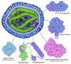
Computer model of cell membrane
2636
A computer model of the cell membrane, where the plasma membrane is red, endoplasmic reticulum is yellow, and mitochondria are blue. Bridget Wilson, University of New Mexico View Media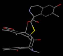
Anti-tumor drug ecteinascidin 743 (ET-743), structure without hydrogens 02
2795
Ecteinascidin 743 (ET-743, brand name Yondelis), was discovered and isolated from a sea squirt, Ecteinascidia turbinata, by NIGMS grantee Kenneth Rinehart at the University of Illinois. Timothy Jamison, Massachusetts Institute of Technology View Media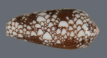
Cone snail shell
2576
A shell from the venomous cone snail Conus omaria, which lives in the Pacific and Indian oceans and eats other snails. Kerry Matz, University of Utah View Media
Structure of a key antigen protein involved with Hepatitis C Virus infection
5866
A three-dimensional representation of the structure of E2, a key antigen protein involved with hepatitis C virus infection. Mansun Law Associate Professor Department of Immunolgy and Microbial Science The Scripps Research Institute View Media
Dimeric ferredoxin-like protein from an unidentified marine microbe
2340
This is the first structure of a protein derived from the metagenomic sequences collected during the Sorcerer II Global Ocean Sampling project. Joint Center for Structural Genomics View Media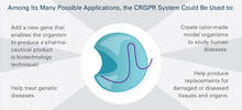
CRISPR Illustration Frame 5
6489
This illustration shows, in simplified terms, how the CRISPR-Cas9 system can be used as a gene-editing tool. This is the fifthframe in a series of five. View Media
Antitoxin GhoS (Illustration 1)
3427
Structure of the bacterial antitoxin protein GhoS. GhoS inhibits the production of a bacterial toxin, GhoT, which can contribute to antibiotic resistance. Rebecca Page and Wolfgang Peti, Brown University and Thomas K. Wood, Pennsylvania State University View Media
Mouse brain 2
6930
A mouse brain that was genetically modified so that subpopulations of its neurons glow. Prayag Murawala, MDI Biological Laboratory and Hannover Medical School. View Media
Bacteria working to eat
2304
Gram-negative bacteria perform molecular acrobatics just to eat. Because they're encased by two membranes, they must haul nutrients across both. Emad Tajkhorshid, University of Illinois at Urbana-Champaign View Media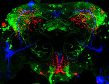
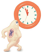
Circadian rhythm neurons in the fruit fly brain
3754
Some nerve cells (neurons) in the brain keep track of the daily cycle. This time-keeping mechanism, called the circadian clock, is found in all animals including us. Justin Blau, New York University View Media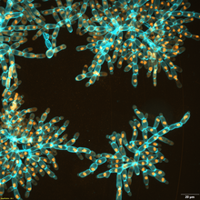
Snowflake yeast 3
6971
Multicellular yeast called snowflake yeast that researchers created through many generations of directed evolution from unicellular yeast. William Ratcliff, Georgia Institute of Technology. View Media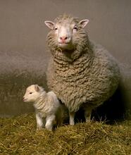
Dolly the sheep
2690
Scientists in Scotland were the first to clone an animal, this sheep named Dolly. She later gave birth to Bonnie, the lamb next to her. View Media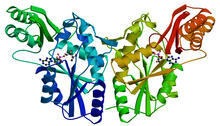
PanC from M. tuberculosis
2383
Model of an enzyme, PanC, that is involved in the last step of vitamin B5 biosynthesis in Mycobacterium tuberculosis. PanC is essential for the growth of M. Mycobacterium Tuberculosis Center, PSI View Media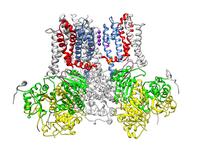
Ion channel
3487
A special "messy" region of a potassium ion channel is important in its function. Yu Zhoi, Christopher Lingle Laboratory, Washington University School of Medicine in St. Louis View Media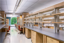
iPS cell facility at the Coriell Institute for Medical Research
2723
This lab space was designed for work on the induced pluripotent stem (iPS) cell collection, part of the NIGMS Human Genetic Cell Repository at the Coriell Institute for Medical Research. Courtney Sill, Coriell Institute for Medical Research View Media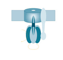
ATP synthase
2517
The world's smallest motor, ATP synthase, generates energy for the cell. See image 2518 for a labeled version of this illustration. Crabtree + Company View Media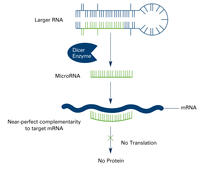
Dicer generates microRNAs (with labels)
2557
The enzyme Dicer generates microRNAs by chopping larger RNA molecules into tiny Velcro®-like pieces. MicroRNAs stick to mRNA molecules and prevent the mRNAs from being made into proteins. Crabtree + Company View Media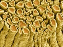
Myelinated axons 1
3396
Myelinated axons in a rat spinal root. Tom Deerinck, National Center for Microscopy and Imaging Research (NCMIR) View Media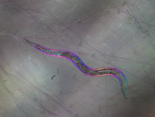
C. elegans trapped by carnivorous fungus
6963
Real-time footage of Caenorhabditis elegans, a tiny roundworm, trapped by a carnivorous fungus, Arthrobotrys dactyloides. Michael Shribak, Marine Biological Laboratory/University of Chicago. View Media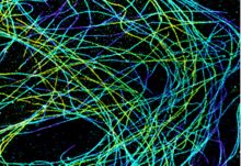
Microtubules in African green monkey cells
6891
Microtubules in African green monkey cells. Microtubules are strong, hollow fibers that provide cells with structural support. Melike Lakadamyali, Perelman School of Medicine at the University of Pennsylvania. View Media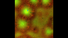
Cell-like compartments emerging from scrambled frog eggs 4
6590
Cell-like compartments that spontaneously emerged from scrambled frog eggs, with nuclei (blue) from frog sperm. Endoplasmic reticulum (red) and microtubules (green) are also visible. Xianrui Cheng, Stanford University School of Medicine. View Media
Diversity oriented synthesis: generating skeletal diversity using folding processes
3327
This 1 1/2-minute video animation was produced for chemical biologist Stuart Schreiber's lab page. The animation shows how diverse chemical structures can be produced in the lab. Eric Keller View Media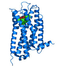
Dopamine D3 receptor
3363
The receptor is shown bound to an antagonist, eticlopride Raymond Stevens, The Scripps Research Institute View Media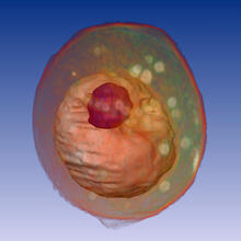
Yeast cell
1092
A whole yeast (Saccharomyces cerevisiae) cell viewed by X-ray microscopy. Inside, the nucleus and a large vacuole (red) are visible. Carolyn Larabell, University of California, San Francisco and the Lawrence Berkeley National Laboratory View Media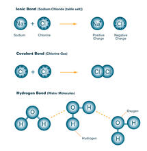
Bond types (with labels)
2520
Ionic and covalent bonds hold molecules, like sodium chloride and chlorine gas, together. Hydrogen bonds among molecules, notably involving water, also play an important role in biology. Crabtree + Company View Media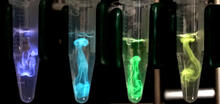
Bioluminescence in a Tube
5895
Details about the basic biology and chemistry of the ingredients that produce bioluminescence are allowing scientists to harness it as an imaging tool. Credit: Nathan Shaner, Scintillon Institute. Nathan Shaner, Scintillon Institute View Media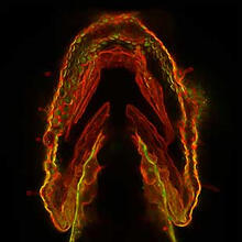
Glowing glycans
2473
Sugars light up the cells in this jaw of a 3-day-old zebrafish embryo and highlight a scientific first: labeling and tracking the movements of sugar chains called glycans in a living organism. Carolyn Bertozzi, University of California, Berkeley View Media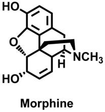
Morphine Structure
3438
The chemical structure of the morphine molecule Judy Coyle, Donald Danforth Plant Science Center View Media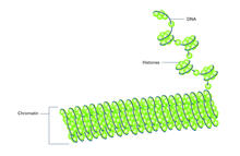
Histones in chromatin (with labels)
2561
Histone proteins loop together with double-stranded DNA to form a structure that resembles beads on a string. Crabtree + Company View Media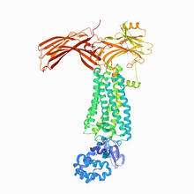
Rhodopsin bound to visual arrestin
6768
Rhodopsin is a pigment in the rod cells of the retina (back of the eye). It is extremely light-sensitive, supporting vision in low-light conditions. Protein Data Bank. View Media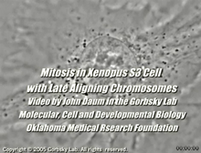
Cell division with late aligning chromosomes
2747
This video shows an instance of abnormal mitosis where chromosomes are late to align. Gary Gorbsky, Oklahoma Medical Research Foundation View Media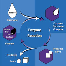
Enzyme reaction
6604
Enzymes speed up chemical reactions by reducing the amount of energy needed for the reactions. NIGMS View Media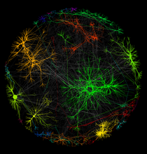
A molecular interaction network in yeast 1
3730
The image visualizes a part of the yeast molecular interaction network. Keiichiro Ono, UCSD View Media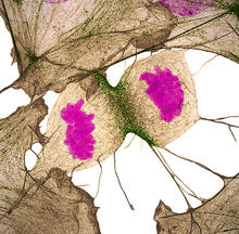
Human fibroblast undergoing cell division
6519
During cell division, cells physically divide after separating their genetic material to create two daughter cells that are genetically identical to the parent cell. Nilay Taneja, Vanderbilt University, and Dylan T. Burnette, Ph.D., Vanderbilt University School of Medicine. View Media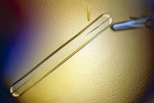
Bovine milk alpha-lactalbumin (1)
2397
A crystal of bovine milk alpha-lactalbumin protein created for X-ray crystallography, which can reveal detailed, three-dimensional protein structures. Alex McPherson, University of California, Irvine View Media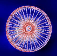
Arachnoidiscus diatom
6902
An Arachnoidiscus diatom with a diameter of 190µm. Michael Shribak, Marine Biological Laboratory/University of Chicago. View Media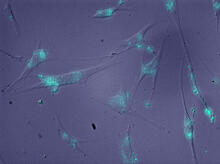
Molecules blocking Huntington's protein production
2600
The molecules that glow blue in these cultured cells prevent the expression of the mutant proteins that cause Huntington's disease. Jiaxin Hu, David W. Dodd and Robert H. E. Hudson, UT Southwestern Medical Center View Media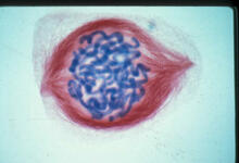
Lily mitosis 05
1015
A light microscope image of a cell from the endosperm of an African globe lily (Scadoxus katherinae). This is one frame of a time-lapse sequence that shows cell division in action. Andrew S. Bajer, University of Oregon, Eugene View Media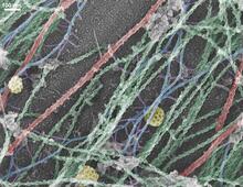
Cells use bubble-like structures called vesicles to transport cargo
3634
Cells use bubble-like structures called vesicles (yellow) to import, transport, and export cargo and in cellular communication. A single cell may be filled with thousands of moving vesicles.Tatyana Svitkina, University of Pennsylvania View Media
Cell cycle
2498
Cells progress through a cycle that consists of phases for growth (blue, green, yellow) and division (red). Cells become quiescent when they exit this cycle (purple). Crabtree + Company View Media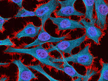
HeLa cells
3521
Multiphoton fluorescence image of HeLa cells stained with the actin binding toxin phalloidin (red), microtubules (cyan) and cell nuclei (blue). Nikon RTS2000MP custom laser scanning microscope. National Center for Microscopy and Imaging Research (NCMIR) View Media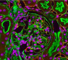
Fluorescent microscopy of kidney tissue--close-up
3725
This photograph of kidney tissue, taken using fluorescent light microscopy, shows a close-up view of part of image 3723. Tom Deerinck , National Center for Microscopy and Imaging Research View Media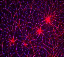
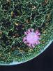
In vitro assembly of a cell-signaling pathway
3787
T cells are white blood cells that are important in defending the body against bacteria, viruses and other pathogens. Xiaolei Su, HHMI Whitman Center of the Marine Biological Laboratory View Media