Switch to Gallery View
Image and Video Gallery
This is a searchable collection of scientific photos, illustrations, and videos. The images and videos in this gallery are licensed under Creative Commons Attribution Non-Commercial ShareAlike 3.0. This license lets you remix, tweak, and build upon this work non-commercially, as long as you credit and license your new creations under identical terms.
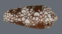
Cone snail shell
2576
A shell from the venomous cone snail Conus omaria, which lives in the Pacific and Indian oceans and eats other snails. Kerry Matz, University of Utah View Media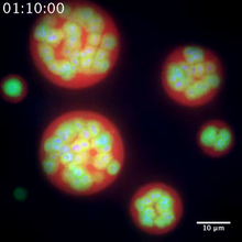
Nucleolus subcompartments spontaneously self-assemble 2
3791
The nucleolus is a small but very important protein complex located in the cell's nucleus. Nilesh Vaidya, Princeton University View Media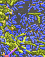
Vibrio bacteria
1160
Vibrio, a type (genus) of rod-shaped bacteria. Some Vibrio species cause cholera in humans. Tina Weatherby Carvalho, University of Hawaii at Manoa View Media
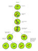
CRISPR Illustration Frame 3
6487
This illustration shows, in simplified terms, how the CRISPR-Cas9 system can be used as a gene-editing tool. National Institute of General Medical Sciences. View Media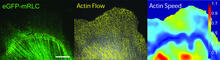
Actin flow
2798
Speckle microscopy analysis of actin cytoskeleton force. This is an example of NIH-supported research on single-cell analysis. Gaudenz Danuser, Harvard Medical School View Media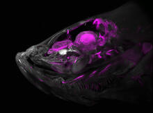
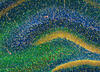
Zebrafish head vasculature
6934
A zebrafish head with blood vessels shown in purple. Prayag Murawala, MDI Biological Laboratory and Hannover Medical School. View Media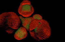
Arabidopsis Thaliana: Flowers Spring to Life
6503
This image capture shows how a single gene, STM, plays a starring role in plant development. Nathanaёl Prunet NIH Support: National Institute of General Medical Sciences View Media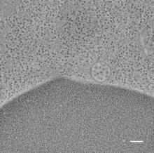
Multivesicular bodies containing intralumenal vesicles assemble at the vacuole 2
5768
Collecting and transporting cellular waste and sorting it into recylable and nonrecylable pieces is a complex business in the cell. Matthew West and Greg Odorizzi, University of Colorado View Media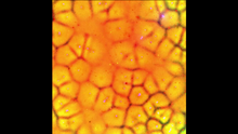
Cell-like compartments emerging from scrambled frog eggs
6587
Cell-like compartments spontaneously emerge from scrambled frog eggs, with nuclei (blue) from frog sperm. Endoplasmic reticulum (red) and microtubules (green) are also visible. Xianrui Cheng, Stanford University School of Medicine. View MediaArtificial cilia exhibit spontaneous beating
3344
Researchers have created artificial cilia that wave like the real thing. Zvonimir Dogic View Media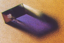
Rabbit GPDA
2405
A crystal of rabbit GPDA protein created for X-ray crystallography, which can reveal detailed, three-dimensional protein structures. Alex McPherson, University of California, Irvine View Media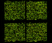
Microarray 01
1070
Microarrays, also called gene chips, are tools that let scientists track the activity of hundreds or thousands of genes simultaneously. Maggie Werner-Washburne, University of New Mexico, Albuquerque View Media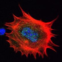
Spreading Cells- 02
3329
Cells move forward with lamellipodia and filopodia supported by networks and bundles of actin filaments. Proper, controlled cell movement is a complex process. Rong Li and Praveen Suraneni, Stowers Institute for Medical Research View Media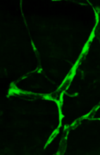
Disrupted and restored vasculature development in frog embryos
3405
Disassembly of vasculature and reassembly after addition and then washout of 250 µM TBZ in kdr:GFP frogs. Hye Ji Cha, University of Texas at Austin View Media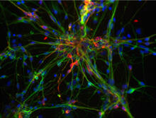
Dopaminergic neurons from ES cells
3270
Human embryonic stem cells differentiated into dopaminergic neurons, the type that degenerate in Parkinson's disease. Image courtesy of the California Institute for Regenerative Medicine. Jeannie Liu, Lab of Jan Nolta, University of California, Davis, via CIRM View Media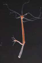
Hydra 01
2437
Hydra magnipapillata is an invertebrate animal used as a model organism to study developmental questions, for example the formation of the body axis. Hiroshi Shimizu, National Institute of Genetics in Mishima, Japan View Media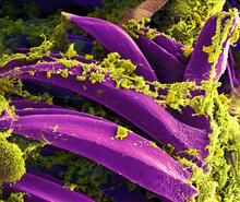
Bubonic plague bacteria on part of the digestive system in a rat flea
3576
Here, bubonic plague bacteria (yellow) are shown in the digestive system of a rat flea (purple). The bubonic plague killed a third of Europeans in the mid-14th century. NIAID View Media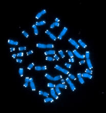
Telomeres
2626
The 46 human chromosomes are shown in blue, with the telomeres appearing as white pinpoints. Hesed Padilla-Nash and Thomas Ried, the National Cancer Institute, a part of NIH View Media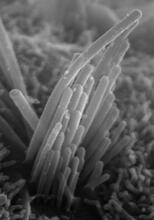
Ear hair cells derived from embryonic stem cells
3272
Mouse embryonic stem cells matured into this bundle of hair cells similar to the ones that transmit sound in the ear. Stefen Heller, Stanford University, via CIRM View MediaTranslation
1281
Ribosomes manufacture proteins based on mRNA instructions. Each ribosome reads mRNA, recruits tRNA molecules to fetch amino acids, and assembles the amino acids in the proper order. Judith Stoffer View Media
Human endoplasmic reticulum membrane protein complex
6777
A 3D model of the human endoplasmic reticulum membrane protein complex (EMC) that identifies its nine essential subunits. Rebecca Voorhees, California Institute of Technology. View Media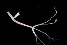
Hydra 06
2442
Hydra magnipapillata is an invertebrate animal used as a model organism to study developmental questions, for example the formation of the body axis. Hiroshi Shimizu, National Institute of Genetics in Mishima, Japan View Media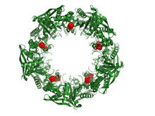
Cas4 nuclease protein structure
3720
This wreath represents the molecular structure of a protein, Cas4, which is part of a system, known as CRISPR, that bacteria use to protect themselves against viral invaders. Fred Dyda, NIDDK View Media
Stretch detectors
2714
Muscles stretch and contract when we walk, and skin splits open and knits back together when we get a paper cut. Christopher Chen, University of Pennsylvania View Media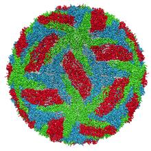
Protective membrane and membrane proteins of the dengue virus visualized with cryo-EM
3756
Dengue virus is a mosquito-borne illness that infects millions of people in the tropics and subtropics each year. Like many viruses, dengue is enclosed by a protective membrane. Hong Zhou, UCLA View Media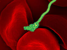
Human blood cells with Borrelia hermsii, a bacterium that causes relapsing fever
3586
Relapsing fever is caused by a bacterium and transmitted by certain soft-bodied ticks or body lice. The disease is seldom fatal in humans, but it can be very serious and prolonged. NIAID View Media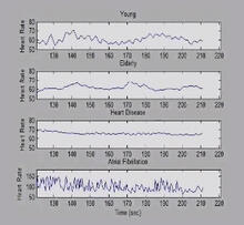
Heart rates time series image
3596
These time series show the heart rates of four different individuals. Madalena Costa and Ary Goldberger, Beth Israel Deaconess Medical Center View Media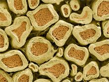
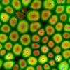
Myelinated axons 2
3397
Top view of myelinated axons in a rat spinal root. Tom Deerinck, National Center for Microscopy and Imaging Research (NCMIR) View Media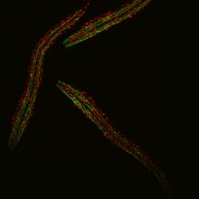
Group of fluorescent C. elegans showing muscle and ribosomal protein
6582
Three C. elegans, tiny roundworms, with a ribosomal protein glowing red and muscle fibers glowing green. Researchers used these worms to study a molecular pathway that affects aging. Jarod Rollins, Mount Desert Island Biological Laboratory. View Media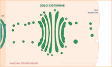
Vesicular shuttle model
1306
Animation for the vesicular shuttle model of Golgi transport. Judith Stoffer View Media
Haplotypes (with labels)
2567
Haplotypes are combinations of gene variants that are likely to be inherited together within the same chromosomal region. Crabtree + Company View Media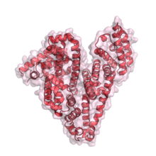
Serum albumin structure 1
3744
Serum albumin (SA) is the most abundant protein in the blood plasma of mammals. SA has a characteristic heart-shape structure and is a highly versatile protein. Wladek Minor, University of Virginia View Media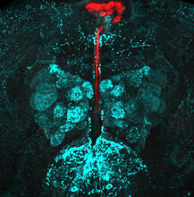
Fruit fly brain responds to adipokines
6985
Drosophila adult brain showing that an adipokine (fat hormone) generates a response from neurons (aqua) and regulates insulin-producing neurons (red).Akhila Rajan, Fred Hutchinson Cancer Center View Media
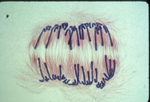
Lily mitosis 10
1010
A light microscope image of a cell from the endosperm of an African globe lily (Scadoxus katherinae). This is one frame of a time-lapse sequence that shows cell division in action. Andrew S. Bajer, University of Oregon, Eugene View Media
Natcher Building 10
1090
NIGMS staff are located in the Natcher Building on the NIH campus. Alisa Machalek, National Institute of General Medical Sciences View Media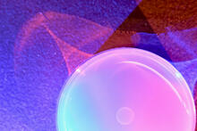
Petri dish
6752
The white circle in this image is a Petri dish, named for its inventor, Julius Richard Petri. H. Robert Horvitz and Dipon Ghosh, Massachusetts Institute of Technology. View Media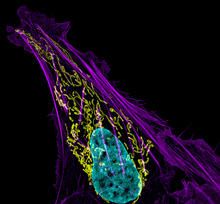
Bone cancer cell
3626
This image shows an osteosarcoma cell with DNA in blue, energy factories (mitochondria) in yellow, and actin filaments—part of the cellular skeleton—in purple. Dylan Burnette and Jennifer Lippincott-Schwartz, NICHD View Media
NMR spectrometer
2371
This photo shows a Varian Unity Inova 900 MHz, 21.1 T standard bore magnet Nuclear Magnetic Resonnance (NMR) spectrometer. Center for Eukaryotic Structural Genomics View Media
Beta2-adrenergic receptor protein
2337
Crystal structure of the beta2-adrenergic receptor protein. The Stevens Laboratory, The Scripps Research Institute View Media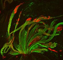
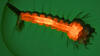
Fruit fly spermatids
3590
Developing spermatids (precursors of mature sperm cells) begin as small, round cells and mature into long-tailed, tadpole-shaped ones. Lacramioara Fabian, The Hospital for Sick Children, Toronto, Canada View Media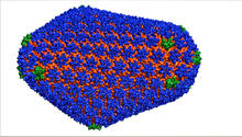
HIV Capsid
3477
This image is a computer-generated model of the approximately 4.2 million atoms of the HIV capsid, the shell that contains the virus' genetic material. Juan R. Perilla and the Theoretical and Computational Biophysics Group, University of Illinois at Urbana-Champaign View Media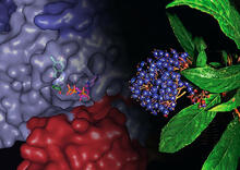
Chang Shan
3483
For thousands of years, Chinese herbalists have treated malaria using Chang Shan, a root extract from a type of hydrangea that grows in Tibet and Nepal. Paul Schimmel Lab, Scripps Research Institute View Media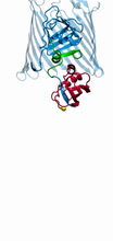
Bacteria working to eat
2304
Gram-negative bacteria perform molecular acrobatics just to eat. Because they're encased by two membranes, they must haul nutrients across both. Emad Tajkhorshid, University of Illinois at Urbana-Champaign View Media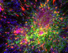
Dopaminergic neurons derived from mouse embryonic stem cells
3271
These neurons are derived from mouse embryonic stem cells. Red shows cells making a protein called TH that is characteristic of the neurons that degenerate in Parkinson's disease. Yaping Sun, lab of Su Guo, University of California, San Francisco, via CIRM View Media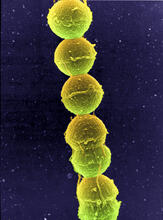
Streptococcus bacteria
1157
Image of Streptococcus, a type (genus) of spherical bacteria that can colonize the throat and back of the mouth. Stroptococci often occur in pairs or in chains, as shown here. Tina Weatherby Carvalho, University of Hawaii at Manoa View Media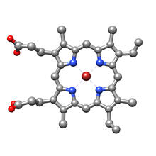
Structure of heme, top view
3539
Molecular model of the struture of heme. Heme is a small, flat molecule with an iron ion (dark red) at its center. Rachel Kramer Green, RCSB Protein Data Bank View Media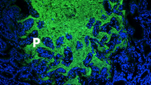
Pathways: What is Basic Science?
6539
Learn about basic science, sometimes called “pure” or “fundamental” science, and how it contributes to the development of medical treatments. National Institute of General Medical Sciences View Media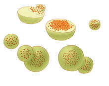
Lysosomes
1282
Lysosomes have powerful enzymes and acids to digest and recycle cell materials. Judith Stoffer View Media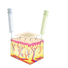
Drugs enter skin
2531
Drugs enter different layers of skin via intramuscular, subcutaneous, or transdermal delivery methods. See image 2532 for a labeled version of this illustration. Crabtree + Company View Media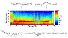
Brain waves of a patient anesthetized with propofol
6779
A representation of a patient’s brain waves after receiving the anesthetic propofol. Emery N. Brown, M.D., Ph.D., Massachusetts General Hospital/Harvard Medical School, Picower Institute for Learning and Memory, and Massachusetts Institute of Technology. View Media