Switch to Gallery View
Image and Video Gallery
This is a searchable collection of scientific photos, illustrations, and videos. The images and videos in this gallery are licensed under Creative Commons Attribution Non-Commercial ShareAlike 3.0. This license lets you remix, tweak, and build upon this work non-commercially, as long as you credit and license your new creations under identical terms.
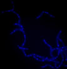
Bacillus anthracis being killed
3525
Bacillus anthracis (anthrax) cells being killed by a fluorescent trans-translation inhibitor, which disrupts bacterial protein synthesis. Kenneth Keiler, Penn State University View Media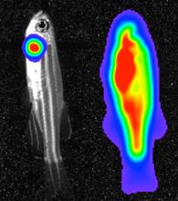
Bioluminescent imaging in adult zebrafish 04
3559
Luciferase-based imaging enables visualization and quantification of internal organs and transplanted cells in live adult zebrafish. View Media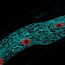
Microsporidia in roundworm 2
5778
Many disease-causing microbes manipulate their host’s metabolism and cells for their own ends. Keir Balla and Emily Troemel, University of California San Diego View Media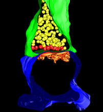
3-D Architecture of a Synapse
5885
This image shows the structure of a synapse, or junction between two nerve cells in three dimensions. From the brain of a mouse. Anton Maximov, The Scripps Research Institute, La Jolla, CA View Media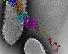
Bacterial nanowire model
6580
A model of a Geobacter sulfurreducens nanowire created from cryo-electron microscopy images. Edward Egelman, University of Virginia. View Media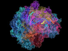
RNA Polymerase II
2484
NIGMS-funded researchers led by Roger Kornberg solved the structure of RNA polymerase II. David Bushnell, Ken Westover and Roger Kornberg, Stanford University View Media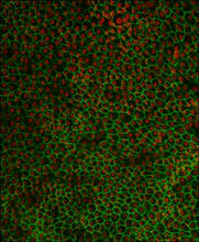
Retinal pigment epithelium derived from human ES cells 02
3287
This image shows a layer of retinal pigment epithelium cells derived from human embryonic stem cells, highlighting the nuclei (red) and cell surfaces (green). David Buckholz and Sherry Hikita, University of California, Santa Barbara, via CIRM View Media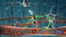
Motion in the brain
2323
Amid a network of blood vessels and star-shaped support cells, neurons in the brain signal each other. The mists of color show the flow of important molecules like glucose and oxygen. Kim Hager and Neal Prakash, University of California, Los Angeles View Media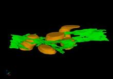
Mitochondria and endoplasmic reticulum
2635
A computer model shows how the endoplasmic reticulum is close to and almost wraps around mitochondria in the cell. The endoplasmic reticulum is lime green and the mitochondria are yellow. Bridget Wilson, University of New Mexico View Media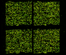
Microarray 01
1070
Microarrays, also called gene chips, are tools that let scientists track the activity of hundreds or thousands of genes simultaneously. Maggie Werner-Washburne, University of New Mexico, Albuquerque View Media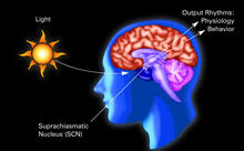
Circadian rhythm (with labels)
2569
The human body keeps time with a master clock called the suprachiasmatic nucleus or SCN. Crabtree + Company View Media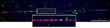
Chromosome fiber 01
2475
This microscopic image shows a chromatin fiber--a DNA molecule bound to naturally occurring proteins. Marc Green and Susan Forsburg, University of Southern California View Media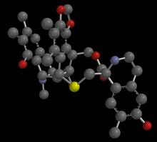
Anti-tumor drug ecteinascidin 743 (ET-743), structure without hydrogens 03
2796
Ecteinascidin 743 (ET-743, brand name Yondelis), was discovered and isolated from a sea squirt, Ecteinascidia turbinata, by NIGMS grantee Kenneth Rinehart at the University of Illinois. Timothy Jamison, Massachusetts Institute of Technology View Media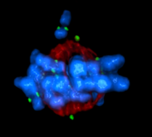
Mitotic cell awaits chromosome alignment
5765
During mitosis, spindle microtubules (red) attach to chromosome pairs (blue), directing them to the spindle equator. View Media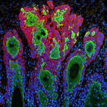
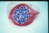
Skin cancer cells (squamous cell carcinoma)
3628
This image shows the uncontrolled growth of cells in squamous cell carcinoma, the second most common form of skin cancer. If caught early, squamous cell carcinoma is usually not life-threatening. Markus Schober and Elaine Fuchs, The Rockefeller University View Media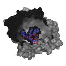
X-ray co-crystal structure of Src kinase bound to a DNA-templated macrocycle inhibitor 3
3415
X-ray co-crystal structure of Src kinase bound to a DNA-templated macrocycle inhibitor. Markus A. Seeliger, Stony Brook University Medical School and David R. Liu, Harvard University View Media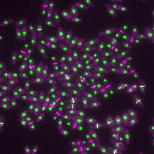
Yeast cells with nuclei and contractile rings
6792
Yeast cells with nuclei shown in green and contractile rings shown in magenta. Nuclei store DNA, and contractile rings help cells divide. Alaina Willet, Kathy Gould’s lab, Vanderbilt University. View Media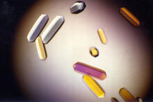
Fungal lipase (1)
2395
Crystals of fungal lipase protein created for X-ray crystallography, which can reveal detailed, three-dimensional protein structures. Alex McPherson, University of California, Irvine View Media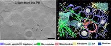
Cryo-ET cell cross-section visualizing insulin vesicles
6607
On the left, a cross-section slice of a rat pancreas cell captured using cryo-electron tomography (cryo-ET). On the right, a color-coded, 3D version of the image highlighting cell structures. Xianjun Zhang, University of Southern California. View Media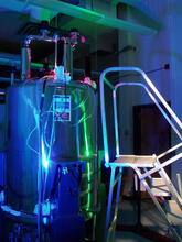
Superconducting magnet
1120
Superconducting magnet for NMR research, from the February 2003 profile of Dorothee Kern in Findings. Mike Lovett View Media
Aldolase
6350
2.5Å resolution reconstruction of rabbit muscle aldolase collected on a FEI/Thermo Fisher Titan Krios with energy filter and image corrector. National Resource for Automated Molecular Microscopy http://nramm.nysbc.org/nramm-images/ Source: Bridget Carragher View Media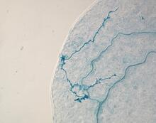
Arabidopsis leaf injected with a pathogen
2780
This is a magnified view of an Arabidopsis thaliana leaf eight days after being infected with the pathogen Hyaloperonospora arabidopsidis, which is closely related to crop pathogens that Jeff Dangl, University of North Carolina, Chapel Hill View Media
Lily mitosis 04
1014
A light microscope image of a cell from the endosperm of an African globe lily (Scadoxus katherinae). This is one frame of a time-lapse sequence that shows cell division in action. Andrew S. Bajer, University of Oregon, Eugene View Media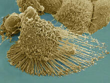
HeLa cells
3519
Scanning electron micrograph of an apoptotic HeLa cell. Zeiss Merlin HR-SEM. National Center for Microscopy and Imaging Research View Media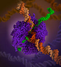
RNA polymerase
6993
RNA polymerase (purple) is a complex enzyme at the heart of transcription. Amy Wu and Christine Zardecki, RCSB Protein Data Bank. View Media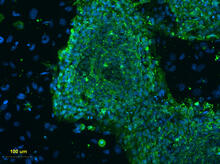
Human embryonic stem cells on feeder cells
3274
This fluorescent microscope image shows human embryonic stem cells whose nuclei are stained green. Blue staining shows the surrounding supportive feeder cells. Michael Longaker lab, Stanford University School of Medicine, via CIRM View Media
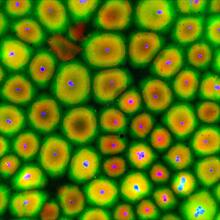
Cell proliferation in a quail embryo
2808
Image showing that the edge zone (top of image) of the quail embryo shows no proliferating cells (cyan), unlike the interior zone (bottom of image). Non-proliferating cell nuclei are labeled green. Andrés Garcia, Georgia Tech View Media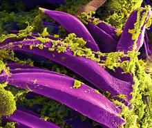
Bubonic plague bacteria on part of the digestive system in a rat flea
3576
Here, bubonic plague bacteria (yellow) are shown in the digestive system of a rat flea (purple). The bubonic plague killed a third of Europeans in the mid-14th century. NIAID View Media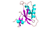
Antitoxin GhoS (Illustration 2)
3428
Structure of the bacterial antitoxin protein GhoS. GhoS inhibits the production of a bacterial toxin, GhoT, which can contribute to antibiotic resistance. Rebecca Page and Wolfgang Peti, Brown University and Thomas K. Wood, Pennsylvania State University View Media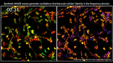
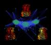
Single-cell “radios” video
7022
Individual cells are color-coded based on their identity and signaling activity using a protein circuit technology developed by the Coyle Lab. Scott Coyle, University of Wisconsin-Madison. View Media
NMR spectrometer
2371
This photo shows a Varian Unity Inova 900 MHz, 21.1 T standard bore magnet Nuclear Magnetic Resonnance (NMR) spectrometer. Center for Eukaryotic Structural Genomics View Media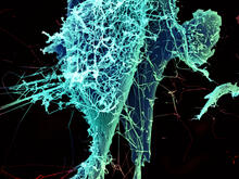
String-like Ebola virus peeling off an infected cell
3619
After multiplying inside a host cell, the stringlike Ebola virus is emerging to infect more cells. Heinz Feldmann, Peter Jahrling, Elizabeth Fischer and Anita Mora, National Institute of Allergy and Infectious Diseases, National Institutes of Health View Media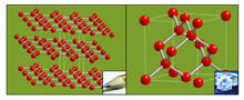
Carbon building blocks (with examples)
2507
The arrangement of identical molecular components can make a dramatic difference. For example, carbon atoms can be arranged into dull graphite (left) or sparkly diamonds (right). Crabtree + Company View Media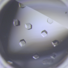
Crystals of CCD-1 in complex with cefotaxime
6764
CCD-1 is an enzyme produced by the bacterium Clostridioides difficile that helps it resist antibiotics. Keith Hodgson, Stanford University. View Media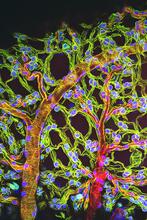
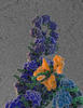
Weblike sheath covering developing egg chambers in a giant grasshopper
3616
The lubber grasshopper, found throughout the southern United States, is frequently used in biology classes to teach students about the respiratory system of insects. Kevin Edwards, Johny Shajahan, and Doug Whitman, Illinois State University. View Media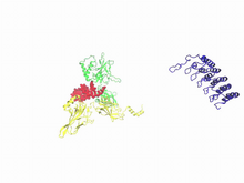
A molecular switch strips transcription factor from DNA
3729
In this video, Rice University scientists used molecular modeling with a mathematical algorithm called AWSEM (for associative memory, water-mediated, structure and energy model) and structural data to Davit Potoyan and Peter Wolynes View Media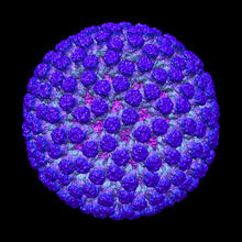
Rotavirus structure
3584
This image shows a computer-generated, three-dimensional map of the rotavirus structure. This virus infects humans and other animals and causes severe diarrhea in infants and young children. Bridget Carragher, The Scripps Research Institute, La Jolla, CA View Media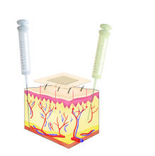
Drugs enter skin
2531
Drugs enter different layers of skin via intramuscular, subcutaneous, or transdermal delivery methods. See image 2532 for a labeled version of this illustration. Crabtree + Company View Media
Human fibroblast undergoing cell division
6519
During cell division, cells physically divide after separating their genetic material to create two daughter cells that are genetically identical to the parent cell. Nilay Taneja, Vanderbilt University, and Dylan T. Burnette, Ph.D., Vanderbilt University School of Medicine. View Media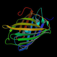
Fruitful dyes
2317
These colorful, computer-generated ribbons show the backbone of a molecule that glows a fluorescent red. Roger Y. Tsien, University of California, San Diego View Media
Rabbit GPDA
2405
A crystal of rabbit GPDA protein created for X-ray crystallography, which can reveal detailed, three-dimensional protein structures. Alex McPherson, University of California, Irvine View Media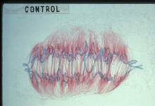
Lily mitosis 09
1022
A light microscope image of a cell from the endosperm of an African globe lily (Scadoxus katherinae). This is one frame of a time-lapse sequence that shows cell division in action. Andrew S. Bajer, University of Oregon, Eugene View Media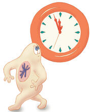
Cell-like compartments from frog eggs
6584
Cell-like compartments that spontaneously emerged from scrambled frog eggs, with nuclei (blue) from frog sperm. Endoplasmic reticulum (red) and microtubules (green) are also visible. Xianrui Cheng, Stanford University School of Medicine. View Media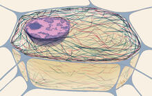
Cytoskeleton
1272
The three fibers of the cytoskeleton--microtubules in blue, intermediate filaments in red, and actin in green--play countless roles in the cell. Judith Stoffer View Media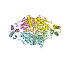
Thymidylate synthase complementing protein from Thermotoga maritime
2387
A model of thymidylate synthase complementing protein from Thermotoga maritime. Joint Center for Structural Genomics, PSI View Media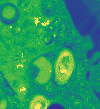
Mycobacterium tuberculosis
2716
Mycobacterium tuberculosis, the bacterium that causes tuberculosis, has infected one-quarter of the world's population and causes more than one million deaths each year, according to the Reuben Peters, Iowa State University View Media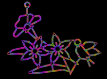
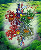
Computer sketch of bird-and-flower DNA origami
3689
A computer-generated sketch of a DNA origami folded into a flower-and-bird structure. See also related image 3690. Hao Yan, Arizona State University View Media
Natcher Building 08
1088
NIGMS staff are located in the Natcher Building on the NIH campus. Alisa Machalek, National Institute of General Medical Sciences View Media
Blood clots show their flex
2450
Blood clots stop bleeding, but they also can cause heart attacks and strokes. Eric Lee, University of Illinois at Urbana-Champaign View Media