
Image Gallery: Lily mitosis 01
ID
1058
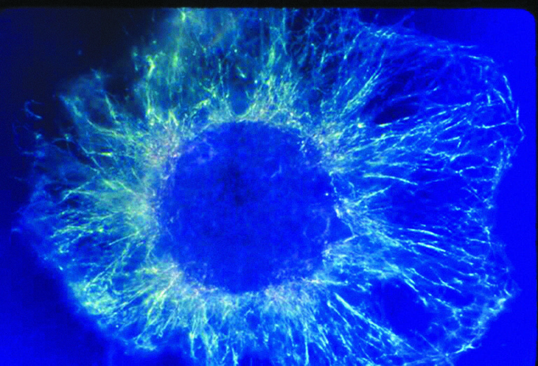
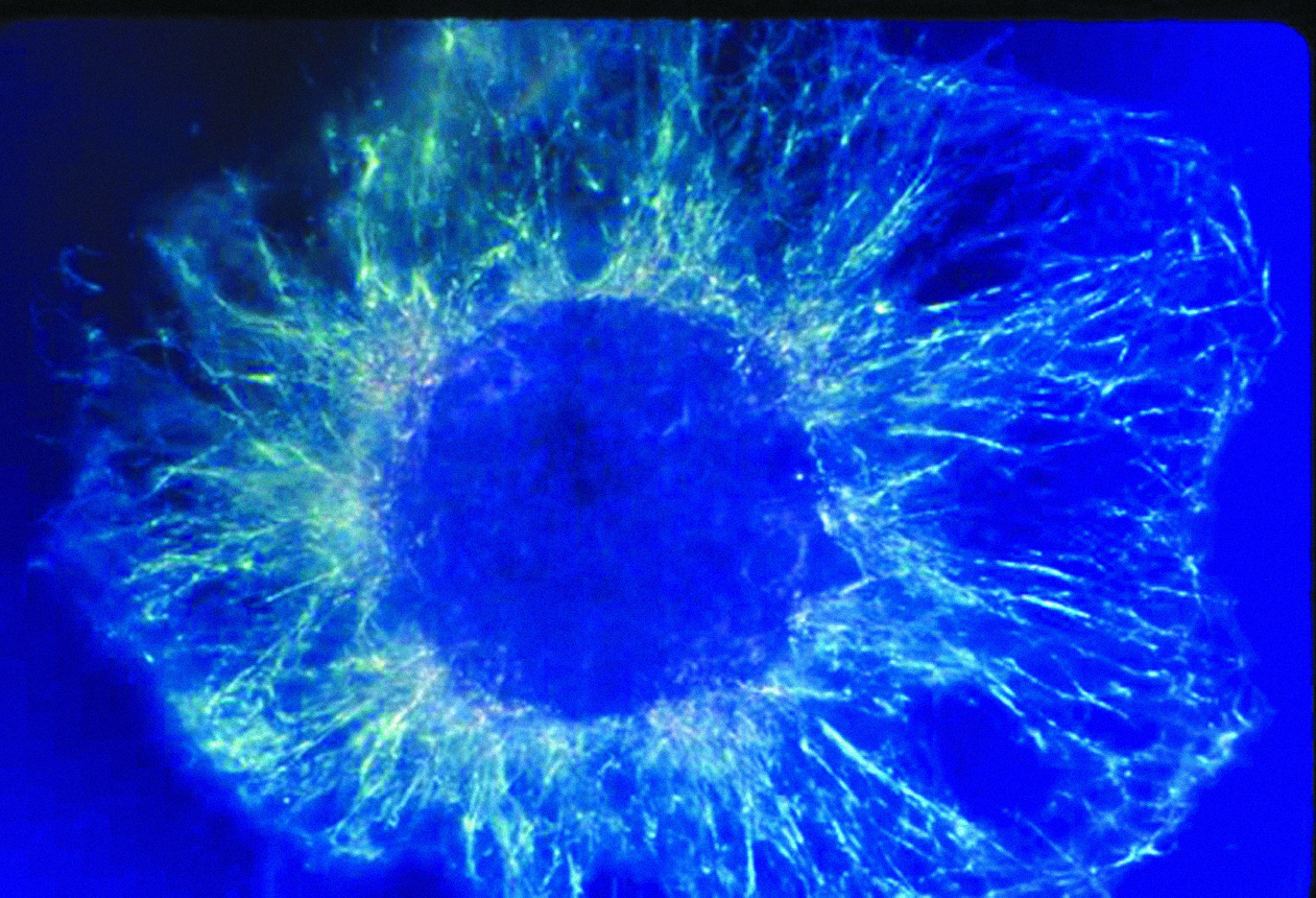
A light microscope image shows the chromosomes, stained dark blue, in a dividing cell of an African globe lily (Scadoxus katherinae). This is one frame of a time-lapse sequence that shows cell division in action. The lily is considered a good organism for studying cell division because its chromosomes are much thicker and easier to see than human ones.
Source
Andrew S. Bajer, University of Oregon, Eugene
