Switch to Gallery View
Image and Video Gallery
This is a searchable collection of scientific photos, illustrations, and videos. The images and videos in this gallery are licensed under Creative Commons Attribution Non-Commercial ShareAlike 3.0. This license lets you remix, tweak, and build upon this work non-commercially, as long as you credit and license your new creations under identical terms.
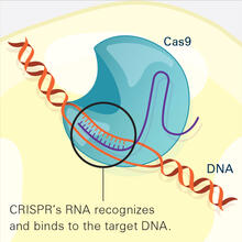
CRISPR Illustration Frame 2
6486
This illustration shows, in simplified terms, how the CRISPR-Cas9 system can be used as a gene-editing tool. National Institute of General Medical Sciences. View Media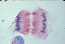
Lily mitosis 11
1011
A light microscope image of cells from the endosperm of an African globe lily (Scadoxus katherinae). This is one frame of a time-lapse sequence that shows cell division in action. Andrew S. Bajer, University of Oregon, Eugene View Media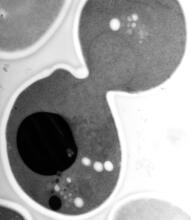
EM of yeast cell division
5770
Cell division is an incredibly coordinated process. Matthew West and Greg Odorizzi, University of Colorado View Media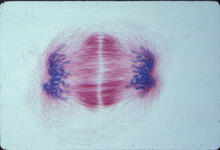
Lily mitosis 12
1018
A light microscope image of a cell from the endosperm of an African globe lily (Scadoxus katherinae). This is one frame of a time-lapse sequence that shows cell division in action. Andrew S. Bajer, University of Oregon, Eugene View Media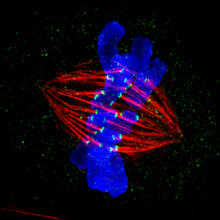
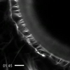
Dividing cell in metaphase
3445
This image of a mammalian epithelial cell, captured in metaphase, was the winning image in the high- and super-resolution microscopy category of the 2012 GE Healthcare Life Sciences Cell Imaging Compe Jane Stout in the laboratory of Claire Walczak, Indiana University, GE Healthcare 2012 Cell Imaging Competition View Media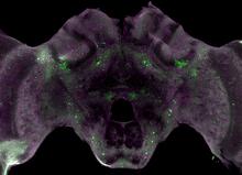
Honeybee brain
6755
Insect brains, like the honeybee brain shown here, are very different in shape from human brains. Gene Robinson, University of Illinois at Urbana-Champaign. View Media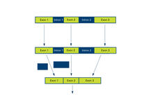
Introns
2550
Genes are often interrupted by stretches of DNA (introns, blue) that do not contain instructions for making a protein. Crabtree + Company View Media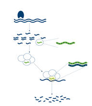
RNA interference
2558
RNA interference or RNAi is a gene-silencing process in which double-stranded RNAs trigger the destruction of specific RNAs. Crabtree + Company View Media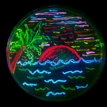
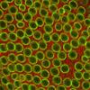
Glowing bacteria make a pretty postcard
3492
This tropical scene, reminiscent of a postcard from Key West, is actually a petri dish containing an artistic arrangement of genetically engineered bacteria. Nathan C. Shaner, The Scintillon Institute View Media
CRISPR Illustration Frame 3
6487
This illustration shows, in simplified terms, how the CRISPR-Cas9 system can be used as a gene-editing tool. National Institute of General Medical Sciences. View Media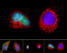
Apoptosis reversed
3486
Two healthy cells (bottom, left) enter into apoptosis (bottom, center) but spring back to life after a fatal toxin is removed (bottom, right; top). Hogan Tang of the Denise Montell Lab, Johns Hopkins University School of Medicine View Media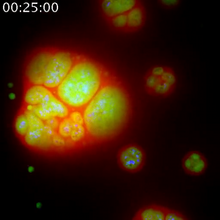
Nucleolus subcompartments spontaneously self-assemble 1
3789
The nucleolus is a small but very important protein complex located in the cell's nucleus. Nilesh Vaidya, Princeton University View Media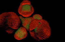
Arabidopsis Thaliana: Flowers Spring to Life
6503
This image capture shows how a single gene, STM, plays a starring role in plant development. Nathanaёl Prunet NIH Support: National Institute of General Medical Sciences View Media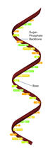
RNA strand (with labels)
2555
Ribonucleic acid (RNA) has a sugar-phosphate backbone and the bases adenine (A), cytosine (C), guanine (G), and uracil (U). Featured in The New Genetics. Crabtree + Company View Media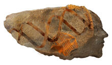
Retroviruses as fossils
2709
DNA doesn't leave a fossil record in stone, the way bones do. Instead, the DNA code itself holds the best evidence for organisms' genetic history. Emily Harrington, science illustrator View Media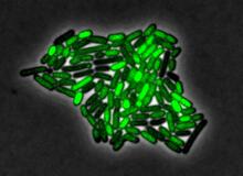
Pulsating response to stress in bacteria
3253
By attaching fluorescent proteins to the genetic circuit responsible for B. subtilis's stress response, researchers can observe the cells' pulses as green flashes. Michael Elowitz, Caltech University View Media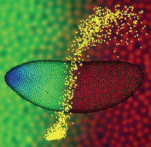
Precise development in the fruit fly embryo
2593
This 2-hour-old fly embryo already has a blueprint for its formation, and the process for following it is so precise that the difference of just a few key molecules can change the plans. Thomas Gregor, Princeton University View Media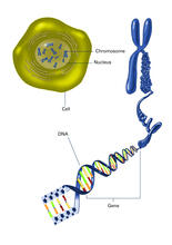
Chromosome inside nucleus (with labels)
2540
The long, stringy DNA that makes up genes is spooled within chromosomes inside the nucleus of a cell. Crabtree + Company View Media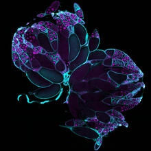
Fruit fly ovaries
6807
Fruit fly (Drosophila melanogaster) ovaries with DNA shown in magenta and actin filaments shown in light blue. This image was captured using a confocal laser scanning microscope.Vladimir I. Gelfand, Feinberg School of Medicine, Northwestern University. View Media
Xenopus laevis embryos
2756
Xenopus laevis, the African clawed frog, has long been used as a model organism for studying embryonic development. The frog embryo on the left lacks the developmental factor Sizzled. Michael Klymkowsky, University of Colorado, Boulder View Media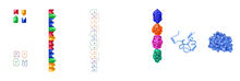
From DNA to Protein
2509
Nucleotides in DNA are copied into RNA, where they are read three at a time to encode the amino acids in a protein. Many parts of a protein fold as the amino acids are strung together. Crabtree + Company View Media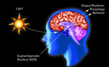
Circadian rhythm (with labels)
2569
The human body keeps time with a master clock called the suprachiasmatic nucleus or SCN. Crabtree + Company View Media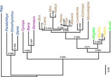
Dinosaur evolutionary tree
2474
Analysis of 68 million-year-old collagen molecule fragments preserved in a T. Chris Organ, Harvard University View Media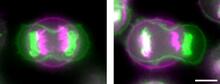
Symmetrically and asymmetrically elongating cells
3648
Merged fluorescent images of symmetrically (left) or asymmetrically (right) elongating HeLa cells at the end of early anaphase (magenta) and late anaphase (green). Tomomi Kiyomitsu and Iain M. Cheeseman, Whitehead Institute for Biomedical Research View Media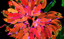
Fruit fly ovary_2
3656
A fruit fly ovary, shown here, contains as many as 20 eggs. Fruit flies are not merely tiny insects that buzz around overripe fruit--they are a venerable scientific tool. Denise Montell, University of California, Santa Barbara View Media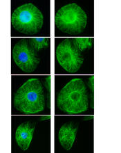
Interphase in Xenopus frog cells
3443
These images show frog cells in interphase. The cells are Xenopus XL177 cells, which are derived from tadpole epithelial cells. The microtubules are green and the chromosomes are blue. Claire Walczak, who took them while working as a postdoc in the laboratory of Timothy Mitchison. View Media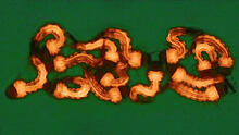
Group of Culex quinquefasciatus mosquito larvae
6770
Mosquito larvae with genes edited by CRISPR. Valentino Gantz, University of California, San Diego. View Media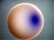
Xenopus laevis egg
2753
Xenopus laevis, the African clawed frog, has long been used as a model organism for studying embryonic development. Michael Klymkowsky, University of Colorado, Boulder View Media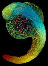
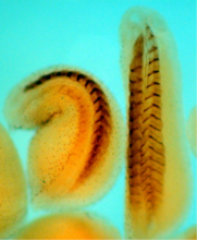
Zebrafish embryo
3644
Just 22 hours after fertilization, this zebrafish embryo is already taking shape. By 36 hours, all of the major organs will have started to form. Philipp Keller, Bill Lemon, Yinan Wan, and Kristin Branson, Janelia Farm Research Campus, Howard Hughes Medical Institute, Ashburn, Va. View Media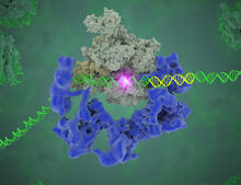
TFIID complex binds DNA to start gene transcription
3766
Gene transcription is a process by which the genetic information encoded in DNA is transcribed into RNA. Eva Nogales, Berkeley Lab View Media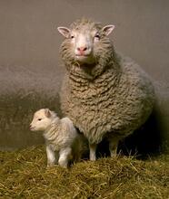
Dolly the sheep
2690
Scientists in Scotland were the first to clone an animal, this sheep named Dolly. She later gave birth to Bonnie, the lamb next to her. View Media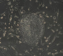
Induced stem cells from adult skin 04
2606
The human skin cells pictured contain genetic modifications that make them pluripotent, essentially equivalent to embryonic stem cells. James Thomson, University of Wisconsin-Madison View Media
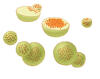
A multicolored fish scale 1
3782
Each of the colored specs in this image is a cell on the surface of a fish scale. Chen-Hui Chen and Kenneth Poss, Duke University View Media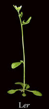
Mature, flowering Arabidopsis
2779
This is an adult flowering Arabidopsis thaliana plant with the inbred designation L-er. Arabidopsis is the most widely used model organism for researchers who study plant genetics. Jeff Dangl, University of North Carolina, Chapel Hill View Media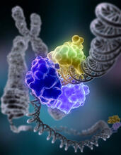
Repairing DNA
2330
Like a watch wrapped around a wrist, a special enzyme encircles the double helix to repair a broken strand of DNA. Tom Ellenberger, Washington University School of Medicine View Media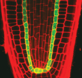
Planting roots
2329
At the root tips of the mustard plant Arabidopsis thaliana (red), two proteins work together to control the uptake of water and nutrients. Philip Benfey, Duke University View Media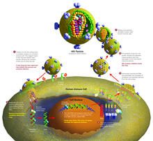
Life of an AIDS virus (with labels and stages)
2515
HIV is a retrovirus, a type of virus that carries its genetic material not as DNA but as RNA. Crabtree + Company View Media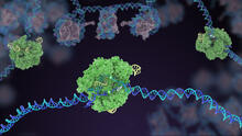
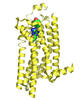
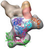
Cas9 protein involved in the CRISPR gene-editing technology
5816
In the gene-editing tool CRISPR, a small strand of RNA identifies a specific chunk of DNA. Janet Iwasa View Media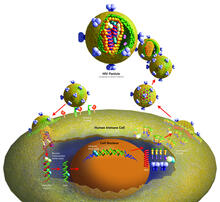
Life of an AIDS virus (with labels)
2514
HIV is a retrovirus, a type of virus that carries its genetic material not as DNA but as RNA. Crabtree + Company View MediaAssembly of the HIV capsid
5729
The HIV capsid is a pear-shaped structure that is made of proteins the virus needs to mature and become infective. John Grime and Gregory Voth, The University of Chicago View Media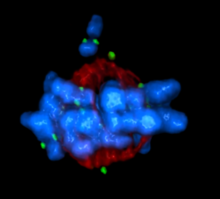
Mitotic cell awaits chromosome alignment
5765
During mitosis, spindle microtubules (red) attach to chromosome pairs (blue), directing them to the spindle equator. View Media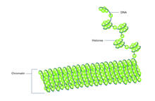
Histones in chromatin (with labels)
2561
Histone proteins loop together with double-stranded DNA to form a structure that resembles beads on a string. Crabtree + Company View Media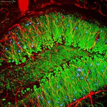
Brain showing hallmarks of Alzheimer's disease
3604
Along with blood vessels (red) and nerve cells (green), this mouse brain shows abnormal protein clumps known as plaques (blue). Alvin Gogineni, Genentech View Media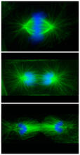
Cell division phases in Xenopus frog cells
3442
These images show three stages of cell division in Xenopus XL177 cells, which are derived from tadpole epithelial cells. They are (from top): metaphase, anaphase and telophase. Claire Walczak, who took them while working as a postdoc in the laboratory of Timothy Mitchison View MediaGenetic mosaicism in fruit flies
6983
Fat tissue from the abdomen of a genetically mosaic adult fruit fly. Genetic mosaicism means that the fly has cells with different genotypes even though it formed from a single zygote. Akhila Rajan, Fred Hutchinson Cancer Center View Media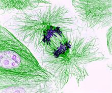
Dividing cells showing chromosomes and cell skeleton
3631
This pig cell is in the process of dividing. The chromosomes (purple) have already replicated and the duplicates are being pulled apart by fibers of the cell skeleton known as microtubules (green). Nasser Rusan, National Heart, Lung, and Blood Institute, National Institutes of Health View Media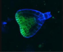
Early development in Arabidopsis
2733
Early on, this Arabidopsis plant embryo picks sides: While one end will form the shoot, the other will take root underground. Zachery R. Smith, Jeff Long lab at the Salk Institute for Biological Studies View Media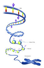
Epigenetic code (with labels)
2563
The "epigenetic code" controls gene activity with chemical tags that mark DNA (purple diamonds) and the "tails" of histone proteins (purple triangles). Crabtree + Company View Media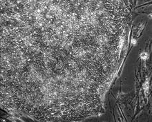
Induced stem cells from adult skin 01
2603
These cells are induced stem cells made from human adult skin cells that were genetically reprogrammed to mimic embryonic stem cells. James Thomson, University of Wisconsin-Madison View Media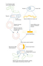
Recombinant DNA (with labels)
2565
To splice a human gene (in this case, the one for insulin) into a plasmid, scientists take the plasmid out of an E. Crabtree + Company View Media