Switch to Gallery View
Image and Video Gallery
This is a searchable collection of scientific photos, illustrations, and videos. The images and videos in this gallery are licensed under Creative Commons Attribution Non-Commercial ShareAlike 3.0. This license lets you remix, tweak, and build upon this work non-commercially, as long as you credit and license your new creations under identical terms.
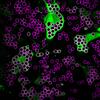
Cell-like compartments emerging from scrambled frog eggs 4
6590
Cell-like compartments that spontaneously emerged from scrambled frog eggs, with nuclei (blue) from frog sperm. Endoplasmic reticulum (red) and microtubules (green) are also visible. Xianrui Cheng, Stanford University School of Medicine. View Media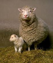
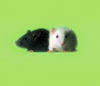
Dolly the sheep
2690
Scientists in Scotland were the first to clone an animal, this sheep named Dolly. She later gave birth to Bonnie, the lamb next to her. View Media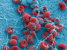
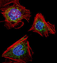
Fibroblasts with nuclei in blue, energy factories in green and the actin cytoskeleton in red
3624
The cells shown here are fibroblasts, one of the most common cells in mammalian connective tissue. These particular cells were taken from a mouse embryo. Dylan Burnette, NICHD View Media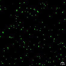
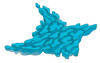
Staphylococcus aureus aggregating upon contact with synovial fluid
6805
Staphylococcus aureus bacteria (green) grouping together upon contact with synovial fluid—a viscous substance found in joints. Paul Stoodley, The Ohio State University. View Media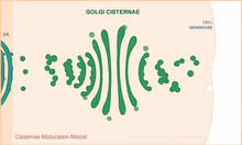
Cisternae maturation model
1307
Animation for the cisternae maturation model of Golgi transport. Judith Stoffer View Media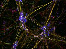
Peripheral nerve cells derived from ES cells
3263
Peripheral nerve cells made from human embryonic stem cell-derived neural crest stem cells. Stephen Dalton, University of Georgia View Media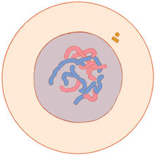
Mitosis - interphase
1316
A cell in interphase, at the start of mitosis: Chromosomes duplicate, and the copies remain attached to each other. Judith Stoffer View Media
Blinking bacteria
2724
Like a pulsing blue shower, E. coli cells flash in synchrony. Genes inserted into each cell turn a fluorescent protein on and off at regular intervals. Jeff Hasty, University of California, San Diego View Media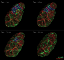
Four timepoints in gastrulation
3334
It has been said that gastrulation is the most important event in a person's life. Bob Goldstein, University of North Carolina, Chapel Hill View Media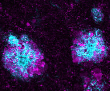
Lysosome clusters around amyloid plaques
5771
It's probably most people's least favorite activity, but we still need to do it--take out our trash. Otherwise our homes will get cluttered and smelly, and eventually, we'll get sick. Swetha Gowrishankar and Shawn Ferguson, Yale School of Medicine View Media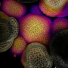
Flower-forming cells in a small plant related to cabbage (Arabidopsis)
3606
In plants, as in animals, stem cells can transform into a variety of different cell types. The stem cells at the growing tip of this Arabidopsis plant will soon become flowers. Arun Sampathkumar and Elliot Meyerowitz, California Institute of Technology View Media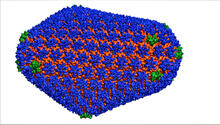
HIV Capsid
3477
This image is a computer-generated model of the approximately 4.2 million atoms of the HIV capsid, the shell that contains the virus' genetic material. Juan R. Perilla and the Theoretical and Computational Biophysics Group, University of Illinois at Urbana-Champaign View Media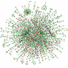
Protein map
2423
Network diagram showing a map of protein-protein interactions in a yeast (Saccharomyces cerevisiae) cell. This cluster includes 78 percent of the proteins in the yeast proteome. Hawoong Jeong, KAIST, Korea View Media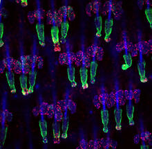
Wound healing in process
3498
Wound healing requires the action of stem cells. Hermann Steller, Rockefeller University View Media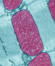
Mitochondria from rat heart muscle cell
3661
These mitochondria (red) are from the heart muscle cell of a rat. Mitochondria have an inner membrane that folds in many places (and that appears here as striations). National Center for Microscopy and Imaging Research View Media
Host infection stimulates antibiotic resistance
5764
This illustration shows pathogenic bacteria behave like a Trojan horse: switching from antibiotic susceptibility to resistance during infection. View Media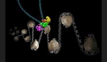
Intasome
6346
Salk researchers captured the structure of a protein complex called an intasome (center) that lets viruses similar to HIV establish permanent infection in their hosts. National Resource for Automated Molecular Microscopy http://nramm.nysbc.org/nramm-images/ Source: Bridget Carragher View Media
Breast cancer cells change migration phenotypes
6986
Cancer cells can change their migration phenotype, which includes their shape and the way that they move to invade different tissues. Bo Sun, Oregon State University. View Media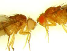
Drosophila
6344
Two adult fruit flies (Drosophila) Dr. Vicki Losick, MDI Biological Laboratory, www.mdibl.org View Media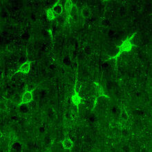
Confocal microscopy of perineuronal nets in the brain 1
3741
The photo shows a confocal microscopy image of perineuronal nets (PNNs), which are specialized extracellular matrix (ECM) structures in the brain. Tom Deerinck, National Center for Microscopy and Imaging Research (NCMIR) View Media
Stress Response in Cells
6570
Two highly stressed osteosarcoma cells are shown with a set of green droplet-like structures followed by a second set of magenta droplets. Julia F. Riley and Carlos A. Castañeda, Syracuse University View Media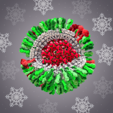
H1N1 Influenza Virus
6355
CellPack image of the H1N1 influenza virus, with hemagglutinin and neuraminidase glycoproteins in green and red, respectively, on the outer envelope (white); matrix protein in gray, and ribonucleoprot Dr. Rommie Amaro, University of California, San Diego View Media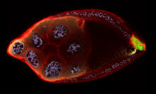
Cellular polarity
2309
As an egg cell develops, a process called polarization controls what parts ultimately become the embryo's head and tail. This picture shows an egg of the fruit fly Drosophila. Wu-Min Deng, Florida State University View Media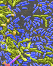
Vibrio bacteria
1160
Vibrio, a type (genus) of rod-shaped bacteria. Some Vibrio species cause cholera in humans. Tina Weatherby Carvalho, University of Hawaii at Manoa View Media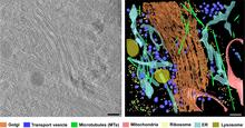
Cryo-ET cross-section of the Golgi apparatus
6606
On the left, a cross-section slice of a rat pancreas cell captured using cryo-electron tomography (cryo-ET). On the right, a 3D, color-coded version of the image highlighting cell structures. Xianjun Zhang, University of Southern California. View Media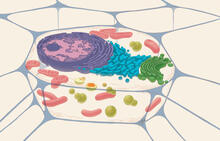
Animal cell
1274
A typical animal cell, sliced open to reveal a cross-section of organelles. Judith Stoffer View Media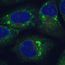
Endoplasmic reticulum abnormalities
6773
Human cells with the gene that codes for the protein FIT2 deleted. Green indicates an endoplasmic reticulum (ER) resident protein. Michel Becuwe, Harvard University. View Media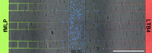
Neutrophil-like cells migrating in a microfluidic chip
6886
Neutrophil-like cells (blue) in a microfluidic chip preferentially migrating toward LTB4 over fMLP. Caroline Jones, University of Texas at Dallas. View Media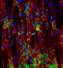
Mouse heart muscle cells
3282
This image shows neonatal mouse heart cells. These cells were grown in the lab on a chip that aligns the cells in a way that mimics what is normally seen in the body. Kara McCloskey lab, University of California, Merced, via CIRM View Media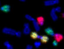
Painted chromosomes
2764
Like a paint-by-numbers picture, painted probes tint individual human chromosomes by targeting specific DNA sequences. Beth A. Sullivan, Duke University View Media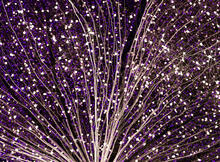
Mouse retina close-up
5872
Keunyoung ("Christine") Kim National Center for Microscopy and Imaging Research (NCMIR) View Media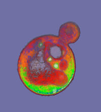
Cells frozen in time
2307
The fledgling field of X-ray microscopy lets researchers look inside whole cells rapidly frozen to capture their actions at that very moment. Here, a yeast cell buds before dividing into two. Carolyn Larabell, University of California, San Francisco, and the Lawrence Berkeley National Laboratory View Media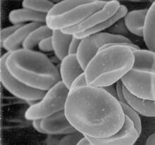
Red blood cells
1101
This image of human red blood cells was obtained with the help of a scanning electron microscope, an instrument that uses a finely focused electron beam to yield detailed images of the surface of a sa Tina Weatherby Carvalho, University of Hawaii at Manoa View Media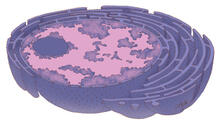
Nucleus and rough ER
1290
The nucleus contains the DNA of eukaryotic cells. Judith Stoffer View Media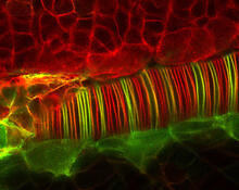
Neural tube development
2328
Proteins in the neural tissues of this zebrafish embryo direct cells to line up and form the neural tube, which will become the spinal cord and brain. Alexander Schier, Harvard University View Media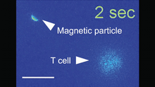
Magnetic Janus particle activating a T cell
6800
A Janus particle being used to activate a T cell, a type of immune cell. Yan Yu, Indiana University, Bloomington. View Media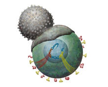
Immune cell attacks cell infected with a retrovirus
2489
T cells engulf and digest cells displaying markers (or antigens) for retroviruses, such as HIV. Kristy Whitehouse, science illustrator View Media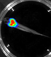
Bioluminescent imaging in adult zebrafish - overhead view
3557
Luciferase-based imaging enables visualization and quantification of internal organs and transplanted cells in live adult zebrafish. Kenneth Poss, Duke University View Media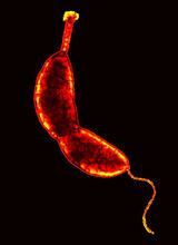
Caulobacter
3262
A study using Caulobacter crescentus showed that some bacteria use just-in-time processing, much like that used in industrial delivery, to make the glue that allows them to attach to surfaces, Yves Brun, Indiana University View Media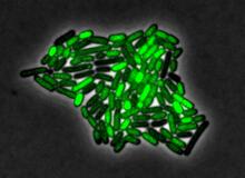
Pulsating response to stress in bacteria
3253
By attaching fluorescent proteins to the genetic circuit responsible for B. subtilis's stress response, researchers can observe the cells' pulses as green flashes. Michael Elowitz, Caltech University View Media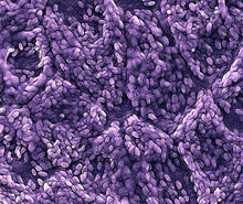
Retinal pigment epithelium derived from human ES cells
3286
This color-enhanced image is a scanning electron microscope image of retinal pigment epithelial (RPE) cells derived from human embryonic stem cells. David Hinton lab, University of Southern California, via CIRM View Media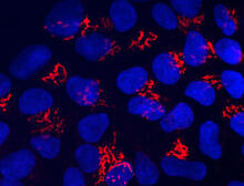
Suicidal Stem Cells
3341
Embryonic stem cells store pre-activated Bax (red) in the Golgi, near the nucleus (blue). Featured in the June 21, 2012, issue of Biomedical Beat. Mohanish Deshmukh View Media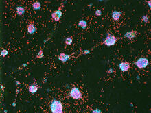
Human ES cells turn into insulin-producing cells
3277
Human embryonic stem cells were differentiated into cells like those found in the pancreas (blue), which give rise to insulin-producing cells (red). Eugene Brandon, ViaCyte, via CIRM View Media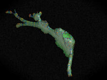
Cell curvature
2803
Rendering of the surface of an endothelial cell; membrane curvature is color coded. This is an example of NIH-supported research on single-cell analysis. Gaudenz Danuser, Harvard Medical School View Media
Floral pattern in a mixture of two bacterial species, Acinetobacter baylyi and Escherichia coli, grown on a semi-solid agar for 72 hour
6556
Floral pattern emerging as two bacterial species, motile Acinetobacter baylyi and non-motile Escherichia coli (green), are grown together for 72 hours on 0.5% agar surface from a small i L. Xiong et al, eLife 2020;9: e48885 View Media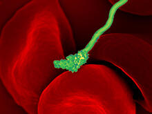
Human blood cells with Borrelia hermsii, a bacterium that causes relapsing fever
3586
Relapsing fever is caused by a bacterium and transmitted by certain soft-bodied ticks or body lice. The disease is seldom fatal in humans, but it can be very serious and prolonged. NIAID View Media
Telomerase illustration
1335
Reactivating telomerase in our cells does not appear to be a good way to extend the human lifespan. Cancer cells reactivate telomerase. Judith Stoffer View Media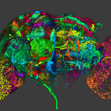
Color coding of the Drosophila brain - image
5838
This image results from a research project to visualize which regions of the adult fruit fly (Drosophila) brain derive from each neural stem cell. Yong Wan from Charles Hansen’s lab, University of Utah. Data preparation and visualization by Masayoshi Ito in the lab of Kei Ito, University of Tokyo. View Media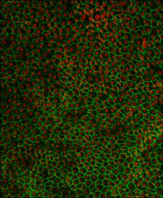
Retinal pigment epithelium derived from human ES cells 02
3287
This image shows a layer of retinal pigment epithelium cells derived from human embryonic stem cells, highlighting the nuclei (red) and cell surfaces (green). David Buckholz and Sherry Hikita, University of California, Santa Barbara, via CIRM View Media