
Image Gallery: Lily mitosis 09
ID
1022
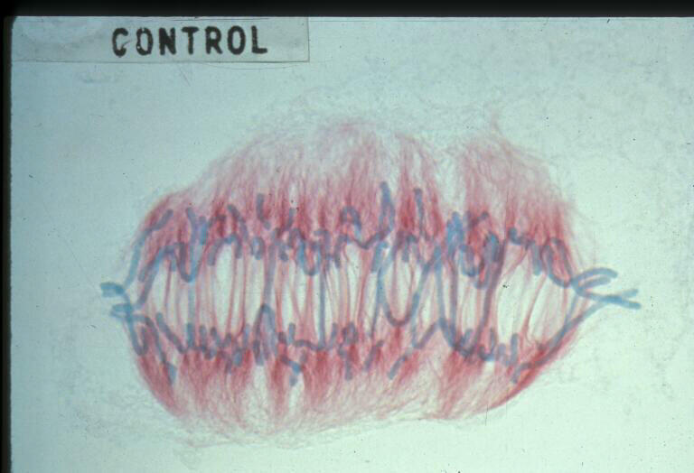
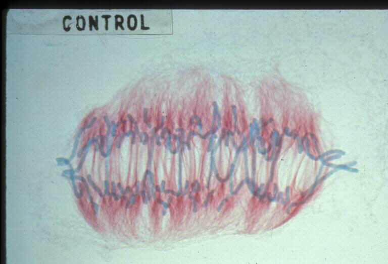
A light microscope image of a cell from the endosperm of an African globe lily (Scadoxus katherinae). This is one frame of a time-lapse sequence that shows cell division in action. The lily is considered a good organism for studying cell division because its chromosomes are much thicker and easier to see than human ones. Staining shows microtubules in red and chromosomes in blue. Here, condensed chromosomes are clearly visible and are starting to separate to form two new cells.
Source
Andrew S. Bajer, University of Oregon, Eugene

