Switch to Gallery View
Image and Video Gallery
This is a searchable collection of scientific photos, illustrations, and videos. The images and videos in this gallery are licensed under Creative Commons Attribution Non-Commercial ShareAlike 3.0. This license lets you remix, tweak, and build upon this work non-commercially, as long as you credit and license your new creations under identical terms.
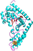
Trp_RS - tryptophanyl tRNA-synthetase family of enzymes
2483
This image represents the structure of TrpRS, a novel member of the tryptophanyl tRNA-synthetase family of enzymes. View Media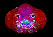
Zebrafish larva
5881
You are face to face with a 6-day-old zebrafish larva. What look like eyes will become nostrils, and the bulges on either side will become eyes. Oscar Ruiz and George Eisenhoffer, University of Texas MD Anderson Cancer Center, Houston View Media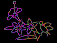
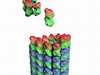
Computer sketch of bird-and-flower DNA origami
3689
A computer-generated sketch of a DNA origami folded into a flower-and-bird structure. See also related image 3690. Hao Yan, Arizona State University View Media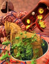
Host infection stimulates antibiotic resistance
5764
This illustration shows pathogenic bacteria behave like a Trojan horse: switching from antibiotic susceptibility to resistance during infection. View Media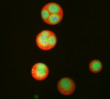
Nucleolus subcompartments spontaneously self-assemble 3
3792
What looks a little like distant planets with some mysterious surface features are actually assemblies of proteins normally found in the cell's nucleolus, a small but very important protein complex lo Nilesh Vaidya, Princeton University View Media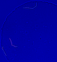
Genetically identical mycobacteria respond differently to antibiotic 2
5752
Antibiotic resistance in microbes is a serious health concern. So researchers have turned their attention to how bacteria undo the action of some antibiotics. Bree Aldridge, Tufts University View Media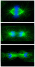
Cell division phases in Xenopus frog cells
3442
These images show three stages of cell division in Xenopus XL177 cells, which are derived from tadpole epithelial cells. They are (from top): metaphase, anaphase and telophase. Claire Walczak, who took them while working as a postdoc in the laboratory of Timothy Mitchison View Media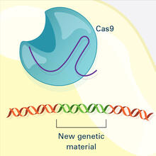
CRISPR Illustration Frame 4
6488
This illustration shows, in simplified terms, how the CRISPR-Cas9 system can be used as a gene-editing tool. National Institute of General Medical Sciences. View Media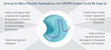
CRISPR Illustration Frame 5
6489
This illustration shows, in simplified terms, how the CRISPR-Cas9 system can be used as a gene-editing tool. This is the fifthframe in a series of five. View Media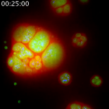
Nucleolus subcompartments spontaneously self-assemble 1
3789
The nucleolus is a small but very important protein complex located in the cell's nucleus. Nilesh Vaidya, Princeton University View Media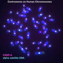
Centromeres on human chromosomes
3255
Human metaphase chromosomes are visible with fluorescence in vitro hybridization (FISH). Centromeric alpha satellite DNA (green) are found in the heterochromatin at each centromere. Peter Warburton, Mount Sinai School of Medicine View Media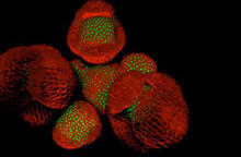
Arabidopsis Thaliana: Flowers Spring to Life
6503
This image capture shows how a single gene, STM, plays a starring role in plant development. Nathanaёl Prunet NIH Support: National Institute of General Medical Sciences View Media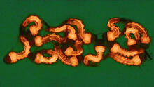
Group of Culex quinquefasciatus mosquito larvae
6770
Mosquito larvae with genes edited by CRISPR. Valentino Gantz, University of California, San Diego. View Media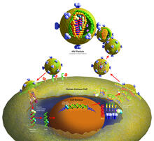
Life of an AIDS virus (with labels)
2514
HIV is a retrovirus, a type of virus that carries its genetic material not as DNA but as RNA. Crabtree + Company View Media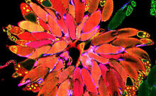
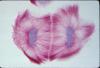
Fruit fly ovary_2
3656
A fruit fly ovary, shown here, contains as many as 20 eggs. Fruit flies are not merely tiny insects that buzz around overripe fruit--they are a venerable scientific tool. Denise Montell, University of California, Santa Barbara View Media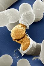
Birth of a yeast cell
3614
Yeast make bread, beer, and wine. And like us, yeast can reproduce sexually. A mother and father cell fuse and create one large cell that contains four offspring. Juergen Berger, Max Planck Institute for Developmental Biology, and Maria Langegger, Friedrich Miescher Laboratory of the Max Planck Society, Germany View Media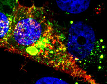
RSV-Infected Cell
3567
Viral RNA (red) in an RSV-infected cell. Eric Alonas and Philip Santangelo, Georgia Institute of Technology and Emory University View Media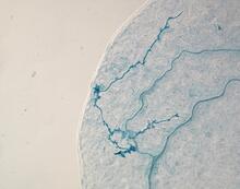
Arabidopsis leaf injected with a pathogen
2780
This is a magnified view of an Arabidopsis thaliana leaf eight days after being infected with the pathogen Hyaloperonospora arabidopsidis, which is closely related to crop pathogens that Jeff Dangl, University of North Carolina, Chapel Hill View Media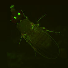
Fly by night
2417
This fruit fly expresses green fluorescent protein (GFP) in the same pattern as the period gene, a gene that regulates circadian rhythm and is expressed in all sensory neurons on the surface of the fl Jay Hirsh, University of Virginia View Media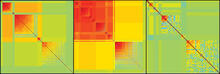
Genetic patchworks
2588
Each point in these colorful patchworks represents the correlation between two sleep-associated genes in fruit flies. Susan Harbison and Trudy Mackay, North Carolina State University View Media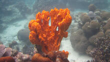
Sponge
2728
Many of today's medicines come from products found in nature, such as this sponge found off the coast of Palau in the Pacific Ocean. Phil Baran, Scripps Research Institute View Media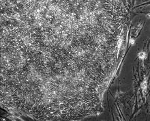
Induced stem cells from adult skin 01
2603
These cells are induced stem cells made from human adult skin cells that were genetically reprogrammed to mimic embryonic stem cells. James Thomson, University of Wisconsin-Madison View Media
Introduction to Genome Editing Using CRISPR/Cas9
5815
Genome editing using CRISPR/Cas9 is a rapidly expanding field of scientific research with emerging applications in disease treatment, medical therapeutics and bioenergy, just to name a few. Janet Iwasa View Media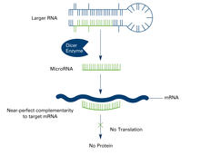
Dicer generates microRNAs (with labels)
2557
The enzyme Dicer generates microRNAs by chopping larger RNA molecules into tiny Velcro®-like pieces. MicroRNAs stick to mRNA molecules and prevent the mRNAs from being made into proteins. Crabtree + Company View Media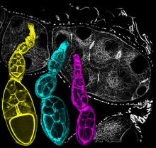
Fruit fly ovarioles
6810
Three fruit fly (Drosophila melanogaster) ovarioles (yellow, blue, and magenta) with egg cells visible inside them. Ovarioles are tubes in the reproductive systems of female insects. Vladimir I. Gelfand, Feinberg School of Medicine, Northwestern University. View Media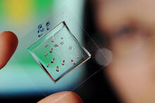
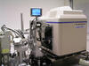
Automated Worm Sorter - 4
3475
Georgia Tech associate professor Hang Lu holds a microfluidic chip that is part of a system that uses artificial intelligence and cutting-edge image processing to automatically examine large number of Georgia Tech/Gary Meek View Media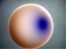
Xenopus laevis egg
2753
Xenopus laevis, the African clawed frog, has long been used as a model organism for studying embryonic development. Michael Klymkowsky, University of Colorado, Boulder View Media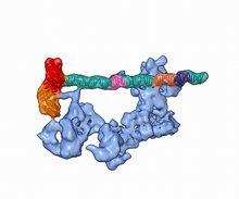
Dynamic cryo-EM model of the human transcription preinitiation complex
5730
Gene transcription is a process by which information encoded in DNA is transcribed into RNA. Eva Nogales, Berkeley Lab View Media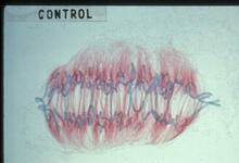
Lily mitosis 09
1022
A light microscope image of a cell from the endosperm of an African globe lily (Scadoxus katherinae). This is one frame of a time-lapse sequence that shows cell division in action. Andrew S. Bajer, University of Oregon, Eugene View Media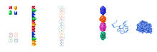
From DNA to Protein
2509
Nucleotides in DNA are copied into RNA, where they are read three at a time to encode the amino acids in a protein. Many parts of a protein fold as the amino acids are strung together. Crabtree + Company View Media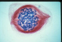
Lily mitosis 05
1015
A light microscope image of a cell from the endosperm of an African globe lily (Scadoxus katherinae). This is one frame of a time-lapse sequence that shows cell division in action. Andrew S. Bajer, University of Oregon, Eugene View Media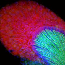
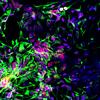
Developing zebrafish fin
3598
Originally from the waters of India, Nepal, and neighboring countries, zebrafish can now be found swimming in science labs (and home aquariums) throughout the world. Jessica Plavicki View Media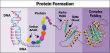
Protein formation
6603
Proteins are 3D structures made up of smaller units. DNA is transcribed to RNA, which in turn is translated into amino acids. NIGMS, with the folded protein illustration adapted from Jane Richardson, Duke University Medical Center View Media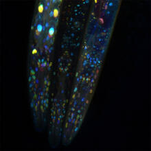
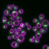
Mosaicism in C. elegans (Black Background)
6532
In the worm C. elegans, double-stranded RNA made in neurons can silence matching genes in a variety of cell types through the transport of RNA between cells. Snusha Ravikumar, Ph.D., University of Maryland, College Park, and Antony M. Jose, Ph.D., University of Maryland, College Park View Media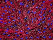
DNA and actin in cultured fibroblast cells
3670
DNA (blue) and actin (red) in cultured fibroblast cells. Tom Deerinck, National Center for Microscopy and Imaging Research (NCMIR) View Media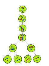
Meiosis illustration
2545
Meiosis is the process whereby a cell reduces its chromosomes from diploid to haploid in creating eggs or sperm. Crabtree + Company View Media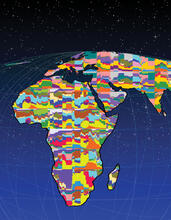
Mapping human genetic variation
2443
This map paints a colorful portrait of human genetic variation around the world. Noah Rosenberg and Martin Soave, University of Michigan View Media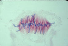
Lily mitosis 07
1017
A light microscope image of a cell from the endosperm of an African globe lily (Scadoxus katherinae). This is one frame of a time-lapse sequence that shows cell division in action. Andrew S. Bajer, University of Oregon, Eugene View Media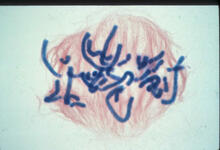
Lily mitosis 06
1016
A light microscope image of a cell from the endosperm of an African globe lily (Scadoxus katherinae). This is one frame of a time-lapse sequence that shows cell division in action. Andrew S. Bajer, University of Oregon, Eugene View Media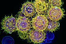
Pollen grains: male germ cells in plants and a cause of seasonal allergies
3609
Those of us who get sneezy and itchy-eyed every spring or fall may have pollen grains, like those shown here, to blame. Edna, Gil, and Amit Cukierman, Fox Chase Cancer Center, Philadelphia, Pa. View Media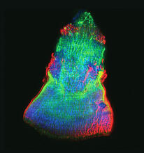
Isolated Planarian Pharynx
3593
The feeding tube, or pharynx, of a planarian worm with cilia shown in red and muscle fibers shown in green View Media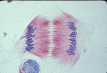
Lily mitosis 11
1011
A light microscope image of cells from the endosperm of an African globe lily (Scadoxus katherinae). This is one frame of a time-lapse sequence that shows cell division in action. Andrew S. Bajer, University of Oregon, Eugene View Media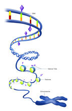
Epigenetic code (with labels)
2563
The "epigenetic code" controls gene activity with chemical tags that mark DNA (purple diamonds) and the "tails" of histone proteins (purple triangles). Crabtree + Company View Media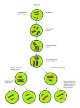
Meiosis illustration (with labels)
2546
Meiosis is the process whereby a cell reduces its chromosomes from diploid to haploid in creating eggs or sperm. Crabtree + Company View Media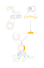
Recombinant DNA
2564
To splice a human gene into a plasmid, scientists take the plasmid out of an E. coli bacterium, cut the plasmid with a restriction enzyme, and splice in human DNA. Crabtree + Company View Media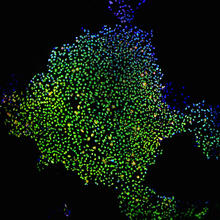
Induced stem cells from adult skin 02
2604
These cells are induced stem cells made from human adult skin cells that were genetically reprogrammed to mimic embryonic stem cells. James Thomson, University of Wisconsin-Madison View Media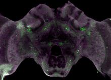
Honeybee brain
6755
Insect brains, like the honeybee brain shown here, are very different in shape from human brains. Gene Robinson, University of Illinois at Urbana-Champaign. View Media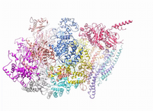
A dynamic model of the DNA helicase protein complex
3750
This short video shows a model of the DNA helicase in yeast. This DNA helicase has 11 proteins that work together to unwind DNA during the process of copying it, called DNA replication. Huilin Li, Stony Brook University View Media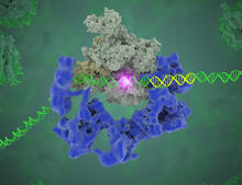
TFIID complex binds DNA to start gene transcription
3766
Gene transcription is a process by which the genetic information encoded in DNA is transcribed into RNA. Eva Nogales, Berkeley Lab View Media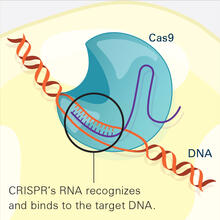
CRISPR Illustration Frame 2
6486
This illustration shows, in simplified terms, how the CRISPR-Cas9 system can be used as a gene-editing tool. National Institute of General Medical Sciences. View Media