Image Gallery: Phagosome in macrophage cell
Video file
ID
6799
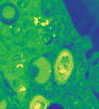
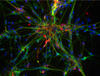
A sensor particle being engulfed by a macrophage—an immune cell—and encapsuled in a compartment called a phagosome. The phagosome then fuses with lysosomes—another type of compartment. The left video shows snowman-shaped sensor particles with fluorescent green nanoparticle “heads” and “bodies” colored red by Förster Resonance Energy Transfer (FRET)-donor fluorophores. The middle video visualizes light blue FRET signals that are only generated when the “snowman” sensor—the FRET-donor—fuses with the lysosomes, which are loaded with FRET-acceptors. The right video combines the other two. The videos were captured using epi-fluorescence microscopy.
More details can be found in the paper “Transport motility of phagosomes on actin and microtubules regulates timing and kinetics of their maturation” by Yu et al.
More details can be found in the paper “Transport motility of phagosomes on actin and microtubules regulates timing and kinetics of their maturation” by Yu et al.
Source
Yan Yu, Indiana University, Bloomington.
Topics