Switch to Gallery View
Image and Video Gallery
This is a searchable collection of scientific photos, illustrations, and videos. The images and videos in this gallery are licensed under Creative Commons Attribution Non-Commercial ShareAlike 3.0. This license lets you remix, tweak, and build upon this work non-commercially, as long as you credit and license your new creations under identical terms.
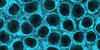
Genetically identical mycobacteria respond differently to antibiotic 2
5752
Antibiotic resistance in microbes is a serious health concern. So researchers have turned their attention to how bacteria undo the action of some antibiotics. Bree Aldridge, Tufts University View Media
Fruit fly spermatids
3590
Developing spermatids (precursors of mature sperm cells) begin as small, round cells and mature into long-tailed, tadpole-shaped ones. Lacramioara Fabian, The Hospital for Sick Children, Toronto, Canada View Media
Interphase in Xenopus frog cells
3443
These images show frog cells in interphase. The cells are Xenopus XL177 cells, which are derived from tadpole epithelial cells. The microtubules are green and the chromosomes are blue. Claire Walczak, who took them while working as a postdoc in the laboratory of Timothy Mitchison. View MediaGenetic mosaicism in fruit flies
6983
Fat tissue from the abdomen of a genetically mosaic adult fruit fly. Genetic mosaicism means that the fly has cells with different genotypes even though it formed from a single zygote. Akhila Rajan, Fred Hutchinson Cancer Center View Media
RNA Polymerase II
2484
NIGMS-funded researchers led by Roger Kornberg solved the structure of RNA polymerase II. David Bushnell, Ken Westover and Roger Kornberg, Stanford University View Media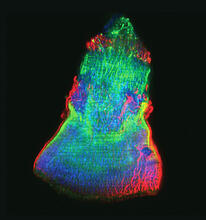
Isolated Planarian Pharynx
3593
The feeding tube, or pharynx, of a planarian worm with cilia shown in red and muscle fibers shown in green View Media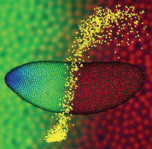
Precise development in the fruit fly embryo
2593
This 2-hour-old fly embryo already has a blueprint for its formation, and the process for following it is so precise that the difference of just a few key molecules can change the plans. Thomas Gregor, Princeton University View MediaCentral dogma, illustrated (with labels)
2548
DNA encodes RNA, which encodes protein. DNA is transcribed to make messenger RNA (mRNA). The mRNA sequence (dark red strand) is complementary to the DNA sequence (blue strand). Crabtree + Company View Media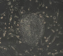
Induced stem cells from adult skin 03
2605
The human skin cells pictured contain genetic modifications that make them pluripotent, essentially equivalent to embryonic stem cells. James Thomson, University of Wisconsin-Madison View MediaAssembly of the HIV capsid
5729
The HIV capsid is a pear-shaped structure that is made of proteins the virus needs to mature and become infective. John Grime and Gregory Voth, The University of Chicago View Media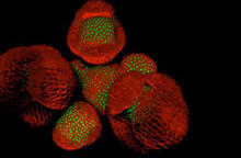
Arabidopsis Thaliana: Flowers Spring to Life
6503
This image capture shows how a single gene, STM, plays a starring role in plant development. Nathanaёl Prunet NIH Support: National Institute of General Medical Sciences View Media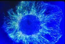
Lily mitosis 01
1058
A light microscope image shows the chromosomes, stained dark blue, in a dividing cell of an African globe lily (Scadoxus katherinae). Andrew S. Bajer, University of Oregon, Eugene View Media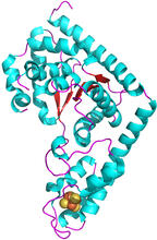
Trp_RS - tryptophanyl tRNA-synthetase family of enzymes
2483
This image represents the structure of TrpRS, a novel member of the tryptophanyl tRNA-synthetase family of enzymes. View Media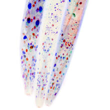
Mosaicism in C. elegans (White Background)
6534
In the worm C. elegans, double-stranded RNA made in neurons can silence matching genes in a variety of cell types through the transport of RNA between cells. Snusha Ravikumar, Ph.D., University of Maryland, College Park, and Antony M. Jose, Ph.D., University of Maryland, College Park View Media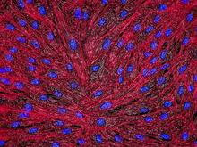
DNA and actin in cultured fibroblast cells
3670
DNA (blue) and actin (red) in cultured fibroblast cells. Tom Deerinck, National Center for Microscopy and Imaging Research (NCMIR) View Media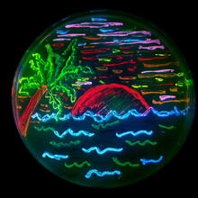
Glowing bacteria make a pretty postcard
3492
This tropical scene, reminiscent of a postcard from Key West, is actually a petri dish containing an artistic arrangement of genetically engineered bacteria. Nathan C. Shaner, The Scintillon Institute View Media
Histones in chromatin
2560
Histone proteins loop together with double-stranded DNA to form a structure that resembles beads on a string. Crabtree + Company View Media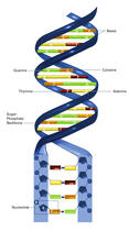
Nucleotides make up DNA (with labels)
2542
DNA consists of two long, twisted chains made up of nucleotides. Each nucleotide contains one base, one phosphate molecule, and the sugar molecule deoxyribose. Crabtree + Company View Media
Induced stem cells from adult skin 02
2604
These cells are induced stem cells made from human adult skin cells that were genetically reprogrammed to mimic embryonic stem cells. James Thomson, University of Wisconsin-Madison View Media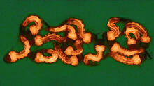
Group of Culex quinquefasciatus mosquito larvae
6770
Mosquito larvae with genes edited by CRISPR. Valentino Gantz, University of California, San Diego. View Media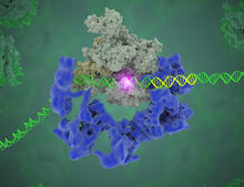
TFIID complex binds DNA to start gene transcription
3766
Gene transcription is a process by which the genetic information encoded in DNA is transcribed into RNA. Eva Nogales, Berkeley Lab View Media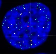
Telomeres on outer edge of nucleus during cell division
3484
New research shows telomeres moving to the outer edge of the nucleus after cell division, suggesting these caps that protect chromosomes also may play a role in organizing DNA. Laure Crabbe, Jamie Kasuboski and James Fitzpatrick, Salk Institute for Biological Studies View Media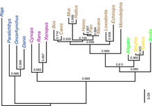
Dinosaur evolutionary tree
2474
Analysis of 68 million-year-old collagen molecule fragments preserved in a T. Chris Organ, Harvard University View Media