Switch to Gallery View
Image and Video Gallery
This is a searchable collection of scientific photos, illustrations, and videos. The images and videos in this gallery are licensed under Creative Commons Attribution Non-Commercial ShareAlike 3.0. This license lets you remix, tweak, and build upon this work non-commercially, as long as you credit and license your new creations under identical terms.
Histones in chromatin
2560
Histone proteins loop together with double-stranded DNA to form a structure that resembles beads on a string. Crabtree + Company View Media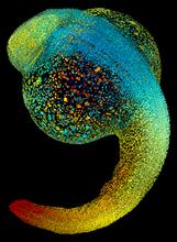
Zebrafish embryo
3644
Just 22 hours after fertilization, this zebrafish embryo is already taking shape. By 36 hours, all of the major organs will have started to form. Philipp Keller, Bill Lemon, Yinan Wan, and Kristin Branson, Janelia Farm Research Campus, Howard Hughes Medical Institute, Ashburn, Va. View Media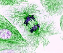
Dividing cells showing chromosomes and cell skeleton
3631
This pig cell is in the process of dividing. The chromosomes (purple) have already replicated and the duplicates are being pulled apart by fibers of the cell skeleton known as microtubules (green). Nasser Rusan, National Heart, Lung, and Blood Institute, National Institutes of Health View Media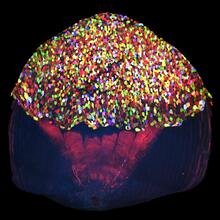
A multicolored fish scale 2
3783
Each of the tiny colored specs in this image is a cell on the surface of a fish scale. Chen-Hui Chen and Kenneth Poss, Duke University View Media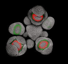
Developing Arabidopsis flower buds
3743
Flower development is a carefully orchestrated, genetically programmed process that ensures that the male (stamen) and female (pistil) organs form in the right place and at the right time in the flowe Nathanaël Prunet, Caltech View Media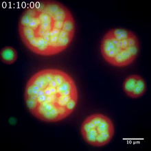
Nucleolus subcompartments spontaneously self-assemble 2
3791
The nucleolus is a small but very important protein complex located in the cell's nucleus. Nilesh Vaidya, Princeton University View Media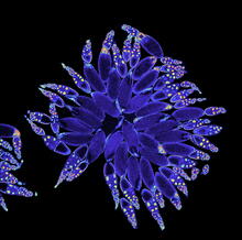
Fruit fly ovary
3607
A fruit fly ovary, shown here, contains as many as 20 eggs. Fruit flies are not merely tiny insects that buzz around overripe fruit—they are a venerable scientific tool. Denise Montell, Johns Hopkins University and University of California, Santa Barbara View Media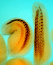
Xenopus laevis embryos
2756
Xenopus laevis, the African clawed frog, has long been used as a model organism for studying embryonic development. The frog embryo on the left lacks the developmental factor Sizzled. Michael Klymkowsky, University of Colorado, Boulder View MediaCentral dogma, illustrated (with labels and numbers for stages)
2549
DNA encodes RNA, which encodes protein. DNA is transcribed to make messenger RNA (mRNA). The mRNA sequence (dark red strand) is complementary to the DNA sequence (blue strand). Crabtree + Company View Media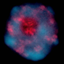
Gene silencing
2318
Pretty in pink, the enzyme histone deacetylase (HDA6) stands out against a background of blue-tinted DNA in the nucleus of an Arabidopsis plant cell. Olga Pontes and Craig Pikaard, Washington University View Media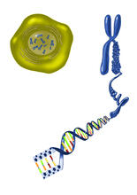
Chromosome inside nucleus
2539
The long, stringy DNA that makes up genes is spooled within chromosomes inside the nucleus of a cell. Crabtree + Company View Media
CRISPR illustration
3719
This illustration shows, in simplified terms, how the CRISPR-Cas9 system can be used as a gene-editing tool. National Institute of General Medical Sciences. View Media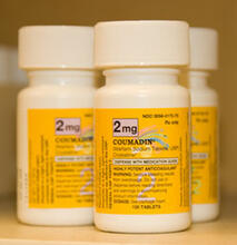
Bottles of warfarin
2579
In 2007, the FDA modified warfarin's label to indicate that genetic makeup may affect patient response to the drug. The widely used blood thinner is sold under the brand name Coumadin®. Alisa Machalek, NIGMS/NIH View MediaCentral dogma, illustrated
2547
DNA encodes RNA, which encodes protein. DNA is transcribed to make messenger RNA (mRNA). The mRNA sequence (dark red strand) is complementary to the DNA sequence (blue strand). Crabtree + Company View Media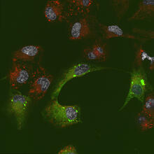
Endoplasmic reticulum abnormalities 2
6774
Human cells with the gene that codes for the protein FIT2 deleted. After an experimental intervention, they are expressing a nonfunctional version of FIT2, shown in green. Michel Becuwe, Harvard University. View Media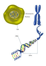
Chromosome inside nucleus (with labels)
2540
The long, stringy DNA that makes up genes is spooled within chromosomes inside the nucleus of a cell. Crabtree + Company View Media
Dicty fruit
2684
Dictyostelium discoideum is a microscopic amoeba. A group of 100,000 form a mound as big as a grain of sand. Featured in The New Genetics. View Media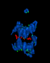
A chromosome goes missing in anaphase
5766
Anaphase is the critical step during mitosis when sister chromosomes are disjoined and directed to opposite spindle poles, ensuring equal distribution of the genome during cell division. View Media
GFP sperm
2683
Fruit fly sperm cells glow bright green when they express the gene for green fluorescent protein (GFP). View Media
Chromosomes before crossing over
1315
Duplicated pair of chromosomes lined up and ready to cross over. Judith Stoffer View Media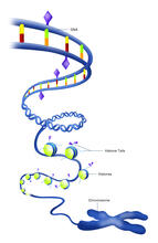
Epigenetic code (with labels)
2563
The "epigenetic code" controls gene activity with chemical tags that mark DNA (purple diamonds) and the "tails" of histone proteins (purple triangles). Crabtree + Company View Media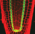
Planting roots
2329
At the root tips of the mustard plant Arabidopsis thaliana (red), two proteins work together to control the uptake of water and nutrients. Philip Benfey, Duke University View Media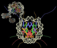
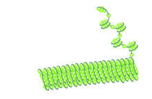
Nucleosome
2741
Like a strand of white pearls, DNA wraps around an assembly of special proteins called histones (colored) to form the nucleosome, a structure responsible for regulating genes and condensing DNA strand Karolin Luger, Colorado State University View Media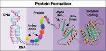
Protein formation
6603
Proteins are 3D structures made up of smaller units. DNA is transcribed to RNA, which in turn is translated into amino acids. NIGMS, with the folded protein illustration adapted from Jane Richardson, Duke University Medical Center View Media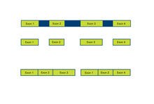
Alternative splicing
2552
Arranging exons in different patterns, called alternative splicing, enables cells to make different proteins from a single gene. Crabtree + Company View Media
Introduction to Genome Editing Using CRISPR/Cas9
5815
Genome editing using CRISPR/Cas9 is a rapidly expanding field of scientific research with emerging applications in disease treatment, medical therapeutics and bioenergy, just to name a few. Janet Iwasa View Media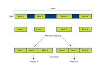
Alternative splicing (with labels)
2553
Arranging exons in different patterns, called alternative splicing, enables cells to make different proteins from a single gene. Featured in The New Genetics. Crabtree + Company View Media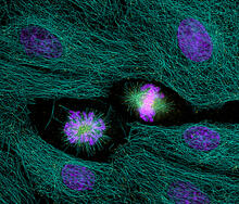
Highlighted cells
2429
The cytoskeleton (green) and DNA (purple) are highlighed in these cells by immunofluorescence. Torsten Wittmann, Scripps Research Institute View Media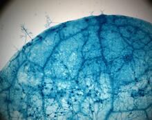
Disease-susceptible Arabidopsis leaf
2782
This is a magnified view of an Arabidopsis thaliana leaf after several days of infection with the pathogen Hyaloperonospora arabidopsidis. Jeff Dangl, University of North Carolina, Chapel Hill View Media
CRISPR Illustration Frame 3
6487
This illustration shows, in simplified terms, how the CRISPR-Cas9 system can be used as a gene-editing tool. National Institute of General Medical Sciences. View Media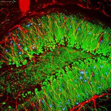
Brain showing hallmarks of Alzheimer's disease
3604
Along with blood vessels (red) and nerve cells (green), this mouse brain shows abnormal protein clumps known as plaques (blue). Alvin Gogineni, Genentech View Media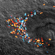
Planarian stem cell colony
3306
Planarians are freshwater flatworms that have powerful abilities to regenerate their bodies, which would seem to make them natural model organisms in which to study stem cells. Peter Reddien, Whitehead Institute View Media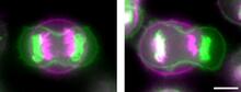
Symmetrically and asymmetrically elongating cells
3648
Merged fluorescent images of symmetrically (left) or asymmetrically (right) elongating HeLa cells at the end of early anaphase (magenta) and late anaphase (green). Tomomi Kiyomitsu and Iain M. Cheeseman, Whitehead Institute for Biomedical Research View Media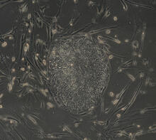
Induced stem cells from adult skin 04
2606
The human skin cells pictured contain genetic modifications that make them pluripotent, essentially equivalent to embryonic stem cells. James Thomson, University of Wisconsin-Madison View Media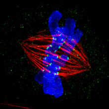
Dividing cell in metaphase
3445
This image of a mammalian epithelial cell, captured in metaphase, was the winning image in the high- and super-resolution microscopy category of the 2012 GE Healthcare Life Sciences Cell Imaging Compe Jane Stout in the laboratory of Claire Walczak, Indiana University, GE Healthcare 2012 Cell Imaging Competition View Media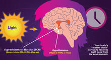
Circadian rhythms and the SCN
6613
Circadian rhythms are physical, mental, and behavioral changes that follow a 24-hour cycle. NIGMS View Media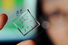
Automated Worm Sorter - 4
3475
Georgia Tech associate professor Hang Lu holds a microfluidic chip that is part of a system that uses artificial intelligence and cutting-edge image processing to automatically examine large number of Georgia Tech/Gary Meek View Media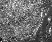
Induced stem cells from adult skin 01
2603
These cells are induced stem cells made from human adult skin cells that were genetically reprogrammed to mimic embryonic stem cells. James Thomson, University of Wisconsin-Madison View Media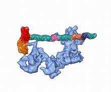
Dynamic cryo-EM model of the human transcription preinitiation complex
5730
Gene transcription is a process by which information encoded in DNA is transcribed into RNA. Eva Nogales, Berkeley Lab View Media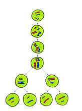
Meiosis illustration
2545
Meiosis is the process whereby a cell reduces its chromosomes from diploid to haploid in creating eggs or sperm. Crabtree + Company View Media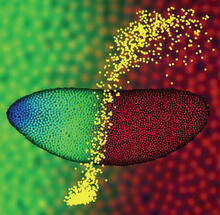
Precise development in the fruit fly embryo
2593
This 2-hour-old fly embryo already has a blueprint for its formation, and the process for following it is so precise that the difference of just a few key molecules can change the plans. Thomas Gregor, Princeton University View MediaAssembly of the HIV capsid
5729
The HIV capsid is a pear-shaped structure that is made of proteins the virus needs to mature and become infective. John Grime and Gregory Voth, The University of Chicago View Media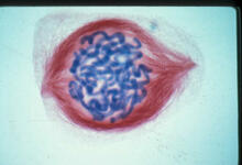
Lily mitosis 05
1015
A light microscope image of a cell from the endosperm of an African globe lily (Scadoxus katherinae). This is one frame of a time-lapse sequence that shows cell division in action. Andrew S. Bajer, University of Oregon, Eugene View Media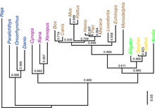
Dinosaur evolutionary tree
2474
Analysis of 68 million-year-old collagen molecule fragments preserved in a T. Chris Organ, Harvard University View Media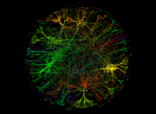
A molecular interaction network in yeast 3
3733
The image visualizes a part of the yeast molecular interaction network. Keiichiro Ono, UCSD View Media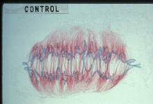
Lily mitosis 09
1022
A light microscope image of a cell from the endosperm of an African globe lily (Scadoxus katherinae). This is one frame of a time-lapse sequence that shows cell division in action. Andrew S. Bajer, University of Oregon, Eugene View Media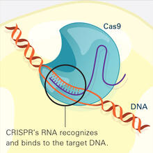
CRISPR Illustration Frame 2
6486
This illustration shows, in simplified terms, how the CRISPR-Cas9 system can be used as a gene-editing tool. National Institute of General Medical Sciences. View Media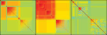
Genetic patchworks
2588
Each point in these colorful patchworks represents the correlation between two sleep-associated genes in fruit flies. Susan Harbison and Trudy Mackay, North Carolina State University View Media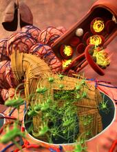
Host infection stimulates antibiotic resistance
5764
This illustration shows pathogenic bacteria behave like a Trojan horse: switching from antibiotic susceptibility to resistance during infection. View Media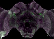
Honeybee brain
6755
Insect brains, like the honeybee brain shown here, are very different in shape from human brains. Gene Robinson, University of Illinois at Urbana-Champaign. View Media