Switch to Gallery View
Image and Video Gallery
This is a searchable collection of scientific photos, illustrations, and videos. The images and videos in this gallery are licensed under Creative Commons Attribution Non-Commercial ShareAlike 3.0. This license lets you remix, tweak, and build upon this work non-commercially, as long as you credit and license your new creations under identical terms.
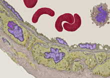
Transmission electron microscopy of coronary artery wall with elastin-rich ECM pseudocolored in light brown
3738
Elastin is a fibrous protein in the extracellular matrix (ECM). It is abundant in artery walls like the one shown here. As its name indicates, elastin confers elasticity. Tom Deerinck, National Center for Microscopy and Imaging Research (NCMIR) View Media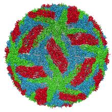
Protective membrane and membrane proteins of the dengue virus visualized with cryo-EM
3756
Dengue virus is a mosquito-borne illness that infects millions of people in the tropics and subtropics each year. Like many viruses, dengue is enclosed by a protective membrane. Hong Zhou, UCLA View Media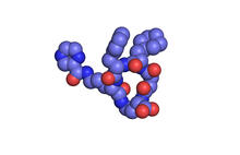
X-ray co-crystal structure of Src kinase bound to a DNA-templated macrocycle inhibitor 1
3413
X-ray co-crystal structure of Src kinase bound to a DNA-templated macrocycle inhibitor. Markus A. Seeliger, Stony Brook University Medical School and David R. Liu, Harvard University View Media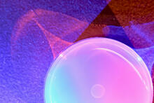
Petri dish
6752
The white circle in this image is a Petri dish, named for its inventor, Julius Richard Petri. H. Robert Horvitz and Dipon Ghosh, Massachusetts Institute of Technology. View Media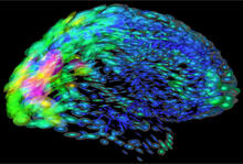
Mapping brain differences
2419
This image of the human brain uses colors and shapes to show neurological differences between two people. Arthur Toga, University of California, Los Angeles View Media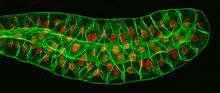
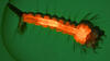
Salivary gland in the developing fruit fly
3603
For fruit flies, the salivary gland is used to secrete materials for making the pupal case, the protective enclosure in which a larva transforms into an adult fly. Richard Fehon, University of Chicago View Media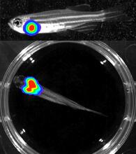
Bioluminescent imaging in adult zebrafish - lateral and overhead view
3556
Luciferase-based imaging enables visualization and quantification of internal organs and transplanted cells in live adult zebrafish. Kenneth Poss, Duke University View Media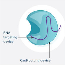
CRISPR Illustration Frame 1
6465
This illustration shows, in simplified terms, how the CRISPR-Cas9 system can be used as a gene-editing tool. This is the first frame in a series of four. National Institute of General Medical Sciences. View Media
Human endoplasmic reticulum membrane protein complex
6777
A 3D model of the human endoplasmic reticulum membrane protein complex (EMC) that identifies its nine essential subunits. Rebecca Voorhees, California Institute of Technology. View Media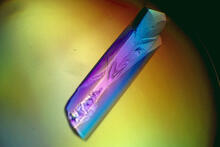
RNase A (1)
2398
A crystal of RNase A protein created for X-ray crystallography, which can reveal detailed, three-dimensional protein structures. Alex McPherson, University of California, Irvine View Media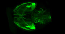
Mouse brain 1
6929
A mouse brain that was genetically modified so that subpopulations of its neurons glow. Prayag Murawala, MDI Biological Laboratory and Hannover Medical School. View Media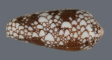
Cone snail shell
2576
A shell from the venomous cone snail Conus omaria, which lives in the Pacific and Indian oceans and eats other snails. Kerry Matz, University of Utah View Media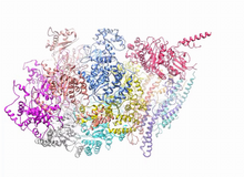
A dynamic model of the DNA helicase protein complex
3750
This short video shows a model of the DNA helicase in yeast. This DNA helicase has 11 proteins that work together to unwind DNA during the process of copying it, called DNA replication. Huilin Li, Stony Brook University View Media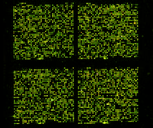
Microarray 01
1070
Microarrays, also called gene chips, are tools that let scientists track the activity of hundreds or thousands of genes simultaneously. Maggie Werner-Washburne, University of New Mexico, Albuquerque View Media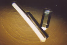
Bovine milk alpha-lactalbumin (2)
2404
Crystals of bovine milk alpha-lactalbumin protein created for X-ray crystallography, which can reveal detailed, three-dimensional protein structures. Alex McPherson, University of California, Irvine View Media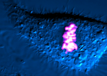
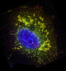
Dividing cell
6965
As this cell was undergoing cell division, it was imaged with two microscopy techniques: differential interference contrast (DIC) and confocal. The DIC view appears in blue and shows the entire cell. Dylan T. Burnette, Vanderbilt University School of Medicine. View Media
Scientists display X-ray diffraction pattern obtained with split X-ray beamline
2384
Scientists from Argonne National Laboratory's Advanced Photon Source (APS) display the first X-ray diffraction pattern obtained from a protein crystal using a split X-ray beam, the first of its kind a GM/CA Collaborative Access Team View Media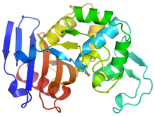
Ribbon diagram of a cefotaxime-CCD-1 complex
6766
CCD-1 is an enzyme produced by the bacterium Clostridioides difficile that helps it resist antibiotics. Keith Hodgson, Stanford University. View Media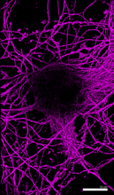
Microtubules in hippocampal neurons
6890
Microtubules (magenta) in neurons of the hippocampus, a part of the brain involved in learning and memory. Microtubules are strong, hollow fibers that provide structural support to cells. Melike Lakadamyali, Perelman School of Medicine at the University of Pennsylvania. View Media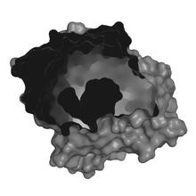
X-ray co-crystal structure of Src kinase bound to a DNA-templated macrocycle inhibitor 2
3414
X-ray co-crystal structure of Src kinase bound to a DNA-templated macrocycle inhibitor. Markus A. Seeliger, Stony Brook University Medical School and David R. Liu, Harvard University View Media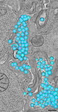
HIV-1 virus in the colon
3571
A tomographic reconstruction of the colon shows the location of large pools of HIV-1 virus particles (in blue) located in the spaces between adjacent cells. Mark Ladinsky, California Institute of Technology View Media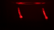
Culex quinquefasciatus mosquito larvae
6771
Mosquito larvae with genes edited by CRISPR swimming in water. Valentino Gantz, University of California, San Diego. View Media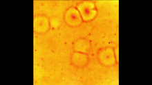
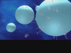
Cell-like compartments emerging from scrambled frog eggs 2
6588
Cell-like compartments spontaneously emerge from scrambled frog eggs, with nuclei (blue) from frog sperm. Endoplasmic reticulum (red) and microtubules (green) are also visible. Xianrui Cheng, Stanford University School of Medicine. View Media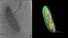
Cryo-electron tomography of a Caulobacter bacterium
6569
3D image of Caulobacter bacterium with various components highlighted: cell membranes (red and blue), protein shell (green), protein factories known as ribosomes (yellow), and storage granules Peter Dahlberg, Stanford University. View Media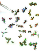
Honeybees marked with paint
6756
Researchers doing behavioral experiments with honeybees sometimes use paint or enamel to give individual bees distinguishing marks. Gene Robinson, University of Illinois at Urbana-Champaign. View Media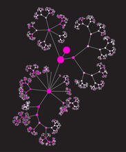
Network diagram of genes, cellular components and processes (unlabeled)
3436
This image shows the hierarchical ontology of genes, cellular components and processes derived from large genomic datasets. From Dutkowski et al. Janusz Dutkowski and Trey Ideker View Media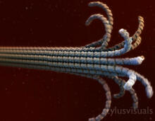
Microtubule breakdown
2321
Like a building supported by a steel frame, a cell contains its own sturdy internal scaffolding made up of proteins, including microtubules. Eva Nogales, University of California, Berkeley View Media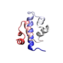
Protein folding video
3391
Proteins are long chains of amino acids. Each protein has a unique amino acid sequence. It is still a mystery how a protein folds into the proper shape based on its sequence. Theoretical and Computational Biophysics Group View Media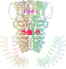
Cryo-electron microscopy revealing the "wasabi receptor"
3747
The TRPA1 protein is responsible for the burn you feel when you taste a bite of sushi topped with wasabi. Jean-Paul Armache, UCSF View Media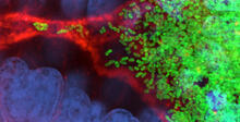
Bacterial cells migrating through the tissues of the squid light organ
7015
Vibrio fischeri cells (~ 2 mm), labeled with green fluorescent protein (GFP), passing through a very narrow bottleneck in the tissues (red) of the Hawaiian bobtail squid, Euprymna scolope Margaret J. McFall-Ngai, Carnegie Institution for Science/California Institute of Technology, and Edward G. Ruby, California Institute of Technology. View Media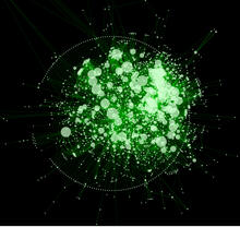
Cytoscape network diagram 1
2737
Molecular biologists are increasingly relying on bioinformatics software to visualize molecular interaction networks and to integrate these networks with data such as gene expression profiles. Keiichiro Ono, Trey Ideker lab, University of California, San Diego View Media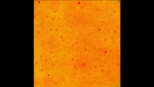
Cell-like compartments emerging from scrambled frog eggs 3
6589
Cell-like compartments spontaneously emerge from scrambled frog eggs. Endoplasmic reticulum (red) and microtubules (green) are visible. Video created using epifluorescence microscopy. Xianrui Cheng, Stanford University School of Medicine. View Media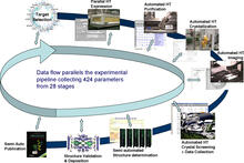
High-throughput protein structure determination pipeline
2364
This slide shows the technologies that the Joint Center for Structural Genomics developed for going from gene to structure and how the technologies have been integrated into a high-throughput pipeline Joint Center for Structural Genomics View Media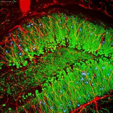
Brain showing hallmarks of Alzheimer's disease
3604
Along with blood vessels (red) and nerve cells (green), this mouse brain shows abnormal protein clumps known as plaques (blue). Alvin Gogineni, Genentech View Media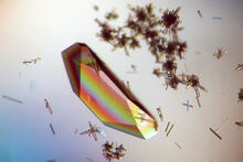
Hen egg lysozyme (1)
2396
Crystals of hen egg lysozyme protein created for X-ray crystallography, which can reveal detailed, three-dimensional protein structures. Alex McPherson, University of California, Irvine View Media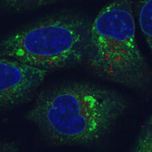
Endoplasmic reticulum abnormalities
6773
Human cells with the gene that codes for the protein FIT2 deleted. Green indicates an endoplasmic reticulum (ER) resident protein. Michel Becuwe, Harvard University. View Media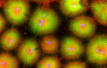
Cell-like compartments from frog eggs 4
6591
Cell-like compartments that spontaneously emerged from scrambled frog eggs, with nuclei (blue) from frog sperm. Endoplasmic reticulum (red) and microtubules (green) are also visible. Xianrui Cheng, Stanford University School of Medicine. View Media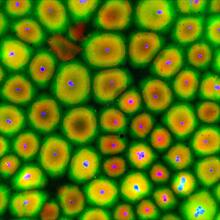
Cell-like compartments from frog eggs
6584
Cell-like compartments that spontaneously emerged from scrambled frog eggs, with nuclei (blue) from frog sperm. Endoplasmic reticulum (red) and microtubules (green) are also visible. Xianrui Cheng, Stanford University School of Medicine. View Media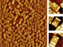
Golden gene chips
2455
A team of chemists and physicists used nanotechnology and DNA's ability to self-assemble with matching RNA to create a new kind of chip for measuring gene activity. Hao Yan and Yonggang Ke, Arizona State University View Media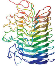
Structure of amyloid-forming prion protein
3542
This structure from an amyloid-forming prion protein shows one way beta sheets can stack. Douglas Fowler, University of Washington View Media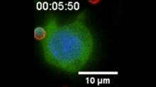
“Two-faced” Janus particle activating a macrophage
6801
A macrophage—a type of immune cell that engulfs invaders—“eats” and is activated by a “two-faced” Janus particle. Yan Yu, Indiana University, Bloomington. View Media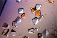
Pig alpha amylase
2412
Crystals of porcine alpha amylase protein created for X-ray crystallography, which can reveal detailed, three-dimensional protein structures. Alex McPherson, University of California, Irvine View Media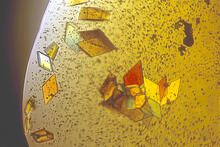
Jack bean concanavalin A
2407
Crystals of jack bean concanavalin A protein created for X-ray crystallography, which can reveal detailed, three-dimensional protein structures. Alex McPherson, University of California, Irvine View Media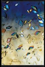
Protein crystals
1060
Structural biologists create crystals of proteins, shown here, as a first step in a process called X-ray crystallography, which can reveal detailed, three-dimensional protein structures. Alex McPherson, University of California, Irvine View Media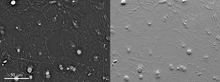
Epithelial cell migration
6899
High-resolution time lapse of epithelial (skin) cell migration and wound healing. It shows an image taken every 13 seconds over the course of almost 14 minutes. Michael Shribak, Marine Biological Laboratory/University of Chicago. View Media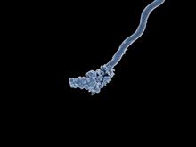
Relapsing fever bacterium (gray) and red blood cells
3585
Relapsing fever is caused by a bacterium and transmitted by certain soft-bodied ticks or body lice. The disease is seldom fatal in humans, but it can be very serious and prolonged. NIAID View Media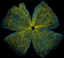
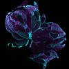
Mouse retina
5793
What looks like the gossamer wings of a butterfly is actually the retina of a mouse, delicately snipped to lay flat and sparkling with fluorescent molecules. Tom Deerinck and Keunyoung (“Christine”) Kim, NCMIR View Media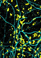
Lysosomes and microtubules
6889
Lysosomes (yellow) and detyrosinated microtubules (light blue). Lysosomes are bubblelike organelles that take in molecules and use enzymes to break them down. Melike Lakadamyali, Perelman School of Medicine at the University of Pennsylvania. View Media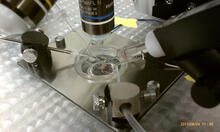
Electrode probe on mouse Huntington's muscle cell
3479
Using an electrode, researchers apply an electrical pulse onto a piece of muscle tissue affected by Huntington's disease. Grigor Varuzhanyan and Andrew A. Voss, California State Polytechnic University View Media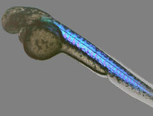
Zebrafish embryo
6897
A zebrafish embryo showing its natural colors. Zebrafish have see-through eggs and embryos, making them ideal research organisms for studying the earliest stages of development. Michael Shribak, Marine Biological Laboratory/University of Chicago. View Media