Switch to Gallery View
Image and Video Gallery
This is a searchable collection of scientific photos, illustrations, and videos. The images and videos in this gallery are licensed under Creative Commons Attribution Non-Commercial ShareAlike 3.0. This license lets you remix, tweak, and build upon this work non-commercially, as long as you credit and license your new creations under identical terms.
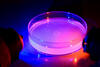
Zebrafish embryo
6897
A zebrafish embryo showing its natural colors. Zebrafish have see-through eggs and embryos, making them ideal research organisms for studying the earliest stages of development. Michael Shribak, Marine Biological Laboratory/University of Chicago. View MediaArtificial cilia exhibit spontaneous beating
3344
Researchers have created artificial cilia that wave like the real thing. Zvonimir Dogic View Media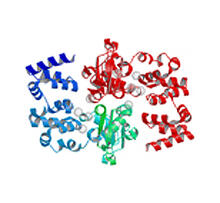
Magnesium transporter protein from E. faecalis
2345
Structure of a magnesium transporter protein from an antibiotic-resistant bacterium (Enterococcus faecalis) found in the human gut. New York Structural GenomiX Consortium View Media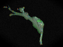
Cell curvature
2803
Rendering of the surface of an endothelial cell; membrane curvature is color coded. This is an example of NIH-supported research on single-cell analysis. Gaudenz Danuser, Harvard Medical School View Media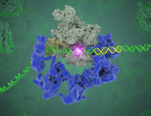
TFIID complex binds DNA to start gene transcription
3766
Gene transcription is a process by which the genetic information encoded in DNA is transcribed into RNA. Eva Nogales, Berkeley Lab View Media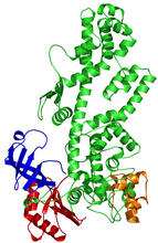
Tex protein
2338
Model of a member from the Tex protein family, which is implicated in transcriptional regulation and highly conserved in eukaryotes and prokaryotes. New York Structural GenomiX Research Consortium, PSI View Media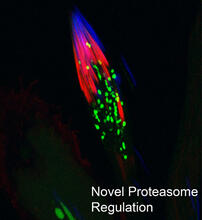
Proteasome
3451
This fruit fly spermatid recycles various molecules, including malformed or damaged proteins. Sigi Benjamin-Hong, Rockefeller University View Media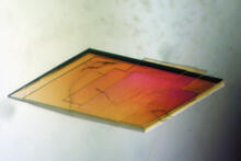
Pig trypsin crystal
2403
A crystal of pig trypsin protein created for X-ray crystallography, which can reveal detailed, three-dimensional protein structures. Alex McPherson, University of California, Irvine View Media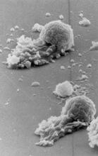
Worm sperm
3489
To develop a system for studying cell motility in unnatrual conditions -- a microscope slide instead of the body -- Tom Roberts and Katsuya Shimabukuro at Florida State University disassembled and rec Tom Roberts, Florida State University View Media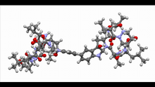
Himastatin, 360-degree view
6851
A 360-degree view of the molecule himastatin, which was first isolated from the bacterium Streptomyces himastatinicus. Himastatin shows antibiotic activity. Mohammad Movassaghi, Massachusetts Institute of Technology. View Media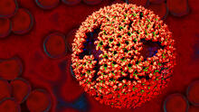
Molecular model of freshly made Rous sarcoma virus (RSV)
3771
Viruses have been the foes of animals and other organisms for time immemorial. Boon Chong Goh, University of Illinois at Urbana-Champaign View Media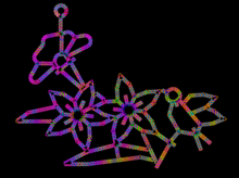
Computer sketch of bird-and-flower DNA origami
3689
A computer-generated sketch of a DNA origami folded into a flower-and-bird structure. See also related image 3690. Hao Yan, Arizona State University View Media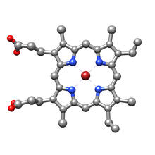
Structure of heme, top view
3539
Molecular model of the struture of heme. Heme is a small, flat molecule with an iron ion (dark red) at its center. Rachel Kramer Green, RCSB Protein Data Bank View Media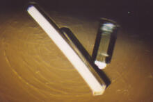
Bovine milk alpha-lactalbumin (2)
2404
Crystals of bovine milk alpha-lactalbumin protein created for X-ray crystallography, which can reveal detailed, three-dimensional protein structures. Alex McPherson, University of California, Irvine View Media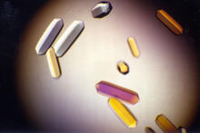
Fungal lipase (1)
2395
Crystals of fungal lipase protein created for X-ray crystallography, which can reveal detailed, three-dimensional protein structures. Alex McPherson, University of California, Irvine View Media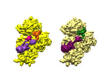
Bacterial ribosome assembly
6578
3D reconstructions of two stages in the assembly of the bacterial ribosome created from time-resolved cryo-electron microscopy images. Ribosomes translate genetic instructions into proteins. Joachim Frank, Columbia University. View Media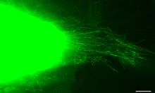
Cytonemes in developing fruit fly cells
3574
Scientists have long known that multicellular organisms use biological molecules produced by one cell and sensed by another to transmit messages that, for instance, guide proper development of organs Sougata Roy, University of California, San Francisco View Media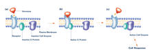
G switch (with labels and stages)
2538
The G switch allows our bodies to respond rapidly to hormones. G proteins act like relay batons to pass messages from circulating hormones into cells. Crabtree + Company View Media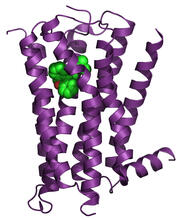
H1 histamine receptor
3360
The receptor is shown bound to an inverse agonist, doxepin. Raymond Stevens, The Scripps Research Institute View Media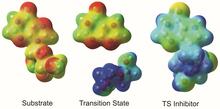
Enzyme transition states
3429
The molecule on the left is an electrostatic potential map of the van der Waals surface of the transition state for human purine nucleoside phosphorylase. Vern Schramm, Albert Einstein College of Medicine of Yeshiva University View Media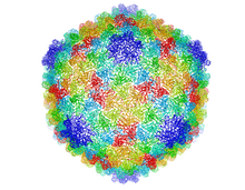
Bacteriophage P22 capsid
5874
Cryo-electron microscopy (cryo-EM) has the power to capture details of proteins and other small biological structures at the molecular level. This image shows proteins in the capsid, or outer co Dr. Wah Chiu, Baylor College of Medicine View Media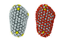
Cryo-EM reveals how the HIV capsid attaches to a human protein to evade immune detection
3755
The illustration shows the capsid of human immunodeficiency virus (HIV) whose molecular features were resolved with cryo-electron microscopy (cryo-EM). Juan R. Perilla, University of Illinois at Urbana-Champaign View Media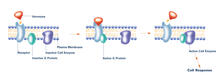
G switch (with labels)
2537
The G switch allows our bodies to respond rapidly to hormones. G proteins act like relay batons to pass messages from circulating hormones into cells. Crabtree + Company View Media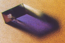
Rabbit GPDA
2405
A crystal of rabbit GPDA protein created for X-ray crystallography, which can reveal detailed, three-dimensional protein structures. Alex McPherson, University of California, Irvine View Media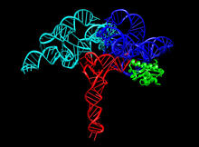
Ribonuclease P structure
3660
Ribbon diagram showing the structure of Ribonuclease P with tRNA. PDB entry 3Q1Q, molecular modeling by Fred Friedman, NIGMS View Media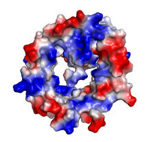
VDAC-1 (4)
2495
The structure of the pore-forming protein VDAC-1 from humans. Gerhard Wagner, Harvard Medical School View Media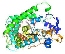
Cytochrome structure with anticancer drug
3326
This image shows the structure of the CYP17A1 enzyme (ribbons colored from blue N-terminus to red C-terminus), with the associated heme colored black. Emily Scott, University of Kansas View Media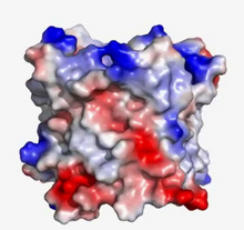
VDAC video 03
2572
This video shows the structure of the pore-forming protein VDAC-1 from humans. Gerhard Wagner, Harvard Medical School View Media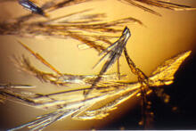
Pig trypsin (3)
2414
Crystals of porcine trypsin protein created for X-ray crystallography, which can reveal detailed, three-dimensional protein structures. Alex McPherson, University of California, Irvine View Media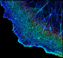
3D image of actin in a cell
3749
Actin is an essential protein in a cell's skeleton (cytoskeleton). It forms a dense network of thin filaments in the cell. Xiaowei Zhuang, Howard Hughes Medical Institute, Harvard University View Media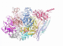
A dynamic model of the DNA helicase protein complex
3750
This short video shows a model of the DNA helicase in yeast. This DNA helicase has 11 proteins that work together to unwind DNA during the process of copying it, called DNA replication. Huilin Li, Stony Brook University View Media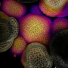
Flower-forming cells in a small plant related to cabbage (Arabidopsis)
3606
In plants, as in animals, stem cells can transform into a variety of different cell types. The stem cells at the growing tip of this Arabidopsis plant will soon become flowers. Arun Sampathkumar and Elliot Meyerowitz, California Institute of Technology View Media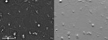
Epithelial cell migration
6899
High-resolution time lapse of epithelial (skin) cell migration and wound healing. It shows an image taken every 13 seconds over the course of almost 14 minutes. Michael Shribak, Marine Biological Laboratory/University of Chicago. View Media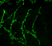
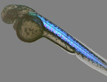
Disrupted vascular development in frog embryos
3403
Disassembly of vasculature in kdr:GFP frogs following addition of 250 µM TBZ. Related to images 3404 and 3505. Hye Ji Cha, University of Texas at Austin View Media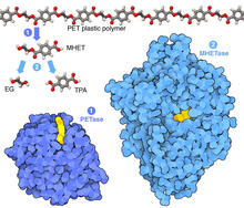
Plastic-eating enzymes
7000
PETase enzyme degrades polyester plastic (polyethylene terephthalate, or PET) into monohydroxyethyl terephthalate (MHET). Amy Wu and Christine Zardecki, RCSB Protein Data Bank. View Media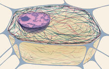
Cytoskeleton
1272
The three fibers of the cytoskeleton--microtubules in blue, intermediate filaments in red, and actin in green--play countless roles in the cell. Judith Stoffer View Media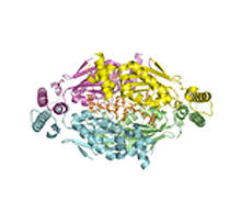
Thymidylate synthase complementing protein from Thermotoga maritime
2387
A model of thymidylate synthase complementing protein from Thermotoga maritime. Joint Center for Structural Genomics, PSI View Media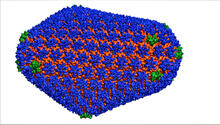
HIV Capsid
3477
This image is a computer-generated model of the approximately 4.2 million atoms of the HIV capsid, the shell that contains the virus' genetic material. Juan R. Perilla and the Theoretical and Computational Biophysics Group, University of Illinois at Urbana-Champaign View Media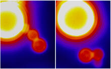
Dynamin Fission
3448
Time lapse series shows short dynamin assemblies (not visible) constricting a lipid tube to make a "beads on a string" appearance, then cutting off one of the beads i.e., catalyzing membrane fission). Ramachandran, Pucadyil et al. , The Scripps Research Institute View Media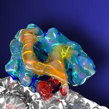
Kinesin moves cellular cargo
3491
A protein called kinesin (blue) is in charge of moving cargo around inside cells and helping them divide. Charles Sindelar, Yale University View Media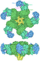
Plant resistosome
7002
The research organism Arabidopsis thaliana forms a large molecular machine called a resistosome to fight off infections. Amy Wu and Christine Zardecki, RCSB Protein Data Bank. View Media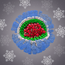
H1N1 Influenza Virus
6356
Related to image 6355. Dr. Rommie Amaro, University of California, San Diego View Media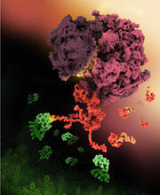
Shiga toxin
6997
E. coli bacteria normally live harmlessly in our intestines, but some cause disease by making toxins. Amy Wu and Christine Zardecki, RCSB Protein Data Bank. View Media
Wreath-shaped protein from X. campestris
2372
Crystal structure of a protein with unknown function from Xanthomonas campestris, a plant pathogen. Eight copies of the protein crystallized to form a ring. Ken Schwinn and Sonia Espejon-Reynes, New York SGX Research Center for Structural Genomics View Media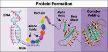
Protein formation
6603
Proteins are 3D structures made up of smaller units. DNA is transcribed to RNA, which in turn is translated into amino acids. NIGMS, with the folded protein illustration adapted from Jane Richardson, Duke University Medical Center View Media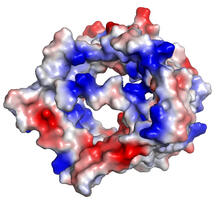
VDAC-1 (2)
2491
The structure of the pore-forming protein VDAC-1 from humans. Gerhard Wagner, Harvard Medical School View Media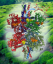
Structure of Glutamate Dehydrogenase
3421
Some children are born with a mutation in a regulatory site on this enzyme that causes them to over-secrete insulin when they consume protein. Judy Coyle, Donald Danforth Plant Science Center View Media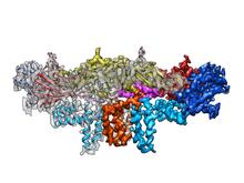
Dengue virus membrane protein structure
3758
Dengue virus is a mosquito-borne illness that infects millions of people in the tropics and subtropics each year. Like many viruses, dengue is enclosed by a protective membrane. Hong Zhou, UCLA View Media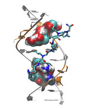
Myotonic dystrophy type 2 genetic defect
3573
Scientists revealed a detailed image of the genetic defect that causes myotonic dystrophy type 2 and used that information to design drug candidates to counteract the disease. Matthew Disney, Scripps Research Institute and Ilyas Yildirim, Northwestern University View Media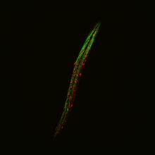
Fluorescent C. elegans showing muscle and ribosomal protein
6581
C. elegans, a tiny roundworm, with a ribosomal protein glowing red and muscle fibers glowing green. Researchers used these worms to study a molecular pathway that affects aging. Jarod Rollins, Mount Desert Island Biological Laboratory. View Media