Switch to Gallery View
Image and Video Gallery
This is a searchable collection of scientific photos, illustrations, and videos. The images and videos in this gallery are licensed under Creative Commons Attribution Non-Commercial ShareAlike 3.0. This license lets you remix, tweak, and build upon this work non-commercially, as long as you credit and license your new creations under identical terms.
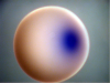
Sphingolipid S1P1 receptor
3362
The receptor is shown bound to an antagonist, ML056. Raymond Stevens, The Scripps Research Institute View Media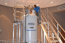
800 MHz NMR magnet
3526
Scientists use nuclear magnetic spectroscopy (NMR) to determine the detailed, 3D structures of molecules. Asokan Anbanandam, University of Kansas View Media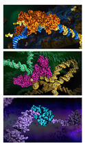
HIV enzyme
6999
These images model the molecular structures of three enzymes with critical roles in the life cycle of the human immunodeficiency virus (HIV). Amy Wu and Christine Zardecki, RCSB Protein Data Bank. View Media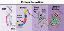
Protein formation
6603
Proteins are 3D structures made up of smaller units. DNA is transcribed to RNA, which in turn is translated into amino acids. NIGMS, with the folded protein illustration adapted from Jane Richardson, Duke University Medical Center View Media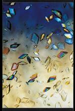
Protein crystals
1060
Structural biologists create crystals of proteins, shown here, as a first step in a process called X-ray crystallography, which can reveal detailed, three-dimensional protein structures. Alex McPherson, University of California, Irvine View Media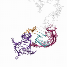
RNA folding in action
6625
An RNA molecule dynamically refolds itself as it is being synthesized. When the RNA is short, it ties itself into a “knot” (dark purple). Julius Lucks, Northwestern University View Media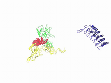
A molecular switch strips transcription factor from DNA
3729
In this video, Rice University scientists used molecular modeling with a mathematical algorithm called AWSEM (for associative memory, water-mediated, structure and energy model) and structural data to Davit Potoyan and Peter Wolynes View Media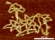
Microscopy image of bird-and-flower DNA origami
3690
An atomic force microscopy image shows DNA folded into an intricate, computer-designed structure. Hao Yan, Arizona State University View Media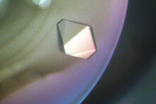
Bacterial glucose isomerase
2409
A crystal of bacterial glucose isomerase protein created for X-ray crystallography, which can reveal detailed, three-dimensional protein structures. Alex McPherson, University of California, Irvine View Media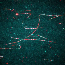
Dynein moving along microtubules
7023
Dynein (green) is a motor protein that “walks” along microtubules (red, part of the cytoskeleton) and carries its cargo along with it. This video was captured through fluorescence microscopy. Morgan DeSantis, University of Michigan. View Media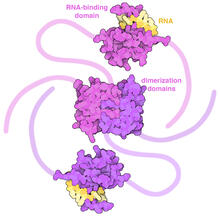
SARS-CoV-2 nucleocapsid dimer
6991
In SARS-CoV-2, the virus that causes COVID-19, nucleocapsid is a complex molecule with many functional parts. Amy Wu and Christine Zardecki, RCSB Protein Data Bank. View Media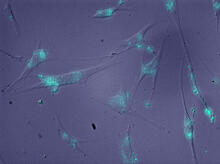
Molecules blocking Huntington's protein production
2600
The molecules that glow blue in these cultured cells prevent the expression of the mutant proteins that cause Huntington's disease. Jiaxin Hu, David W. Dodd and Robert H. E. Hudson, UT Southwestern Medical Center View Media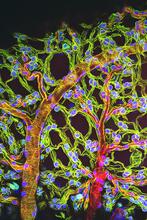
Weblike sheath covering developing egg chambers in a giant grasshopper
3616
The lubber grasshopper, found throughout the southern United States, is frequently used in biology classes to teach students about the respiratory system of insects. Kevin Edwards, Johny Shajahan, and Doug Whitman, Illinois State University. View Media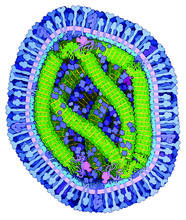
Measles virus
6995
A cross section of the measles virus in which six proteins work together to infect cells. The measles virus is extremely infectious; 9 out of 10 people exposed will contract the disease. Amy Wu and Christine Zardecki, RCSB Protein Data Bank. View Media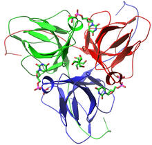
dUTP pyrophosphatase from M. tuberculosis
2381
Model of an enzyme, dUTP pyrophosphatase, from Mycobacterium tuberculosis. Drugs targeted to this enzyme might inhibit the replication of the bacterium that causes most cases of tuberculosis. Mycobacterium Tuberculosis Center, PSI View Media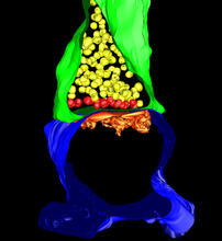
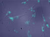
3-D Architecture of a Synapse
5885
This image shows the structure of a synapse, or junction between two nerve cells in three dimensions. From the brain of a mouse. Anton Maximov, The Scripps Research Institute, La Jolla, CA View Media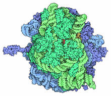
Ribosome illustration from PDB
5780
Ribosomes are complex machines made up of more than 50 proteins and three or four strands of genetic material called ribosomal RNA (rRNA). From PDB’s Molecule of the Month collection (direct link: http://pdb101.rcsb.org/motm/121) Molecule of the Month illustrations are available under a CC-BY-4.0 license. Attribution should be given to David S. Goodsell and the RCSB PDB. View Media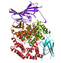
Aminopeptidase N from N. meningitidis
2341
Model of the enzyme aminopeptidase N from the human pathogen Neisseria meningitidis, which can cause meningitis epidemics. Midwest Center for Structural Genomics, PSI View Media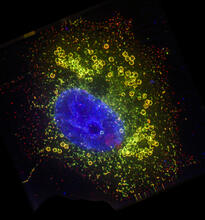
Cell Nucleus and Lipid Droplets
6547
A cell nucleus (blue) surrounded by lipid droplets (yellow). James Olzmann, University of California, Berkeley View Media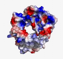
VDAC video 02
2571
This video shows the structure of the pore-forming protein VDAC-1 from humans. Gerhard Wagner, Harvard Medical School View Media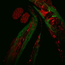
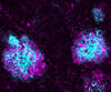
Closeup of fluorescent C. elegans showing muscle and ribosomal protein
6583
Closeup of C. elegans, tiny roundworms, with a ribosomal protein glowing red and muscle fibers glowing green. Researchers used these worms to study a molecular pathway that affects aging. Jarod Rollins, Mount Desert Island Biological Laboratory. View Media
Z rings in bacterial division
2456
Lab-made liposomes contract where Z rings have gathered together and the constriction forces are greatest (arrows). Masaki Osawa, Duke University View Media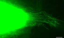
Cytonemes in developing fruit fly cells
3574
Scientists have long known that multicellular organisms use biological molecules produced by one cell and sensed by another to transmit messages that, for instance, guide proper development of organs Sougata Roy, University of California, San Francisco View Media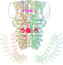
Cryo-electron microscopy revealing the "wasabi receptor"
3747
The TRPA1 protein is responsible for the burn you feel when you taste a bite of sushi topped with wasabi. Jean-Paul Armache, UCSF View MediaNuclear Lamina – Three Views
6573
Three views of the entire nuclear lamina of a HeLa cell produced by tilted light sheet 3D single-molecule super-resolution imaging using a platform termed TILT3D. Anna-Karin Gustavsson, Ph.D. View Media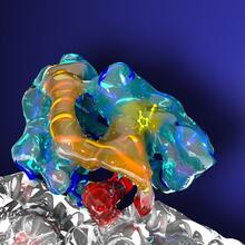
Kinesin moves cellular cargo
3491
A protein called kinesin (blue) is in charge of moving cargo around inside cells and helping them divide. Charles Sindelar, Yale University View Media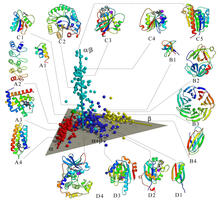
Map of protein structures 02
2367
A global "map of the protein structure universe" indicating the positions of specific proteins. Berkeley Structural Genomics Center, PSI View Media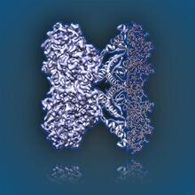
Aldolase
6350
2.5Å resolution reconstruction of rabbit muscle aldolase collected on a FEI/Thermo Fisher Titan Krios with energy filter and image corrector. National Resource for Automated Molecular Microscopy http://nramm.nysbc.org/nramm-images/ Source: Bridget Carragher View Media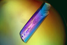
RNase A (1)
2398
A crystal of RNase A protein created for X-ray crystallography, which can reveal detailed, three-dimensional protein structures. Alex McPherson, University of California, Irvine View Media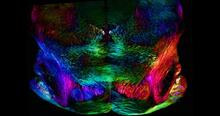
Mouse brain slice showing nerve cells
6901
A 20-µm thick section of mouse midbrain. The nerve cells are transparent and weren’t stained. Michael Shribak, Marine Biological Laboratory/University of Chicago. View Media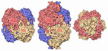
Catalase diversity
7003
Catalases are some of the most efficient enzymes found in cells. Amy Wu and Christine Zardecki, RCSB Protein Data Bank. View Media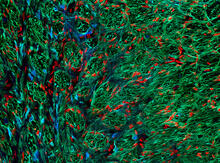
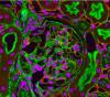
Optic nerve astrocytes
5852
Astrocytes in the cross section of a human optic nerve head Tom Deerinck and Keunyoung (“Christine”) Kim, NCMIR View Media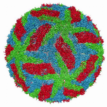
Cryo-electron microscopy of the dengue virus showing protective membrane and membrane proteins
3748
Dengue virus is a mosquito-borne illness that infects millions of people in the tropics and subtropics each year. Like many viruses, dengue is enclosed by a protective membrane. Hong Zhou, UCLA View Media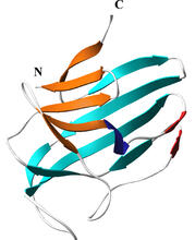
Secreted protein from Mycobacteria
2379
Model of a major secreted protein of unknown function, which is only found in mycobacteria, the class of bacteria that causes tuberculosis. Mycobacterium Tuberculosis Center, PSI View Media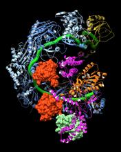
CRISPR surveillance complex
6352
This image shows how the CRISPR surveillance complex is disabled by two copies of anti-CRISPR protein AcrF1 (red) and one AcrF2 (light green). NRAMM National Resource for Automated Molecular Microscopy http://nramm.nysbc.org/nramm-images/ Source: Bridget Carragher View Media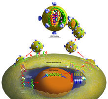
Life of an AIDS virus (with labels)
2514
HIV is a retrovirus, a type of virus that carries its genetic material not as DNA but as RNA. Crabtree + Company View Media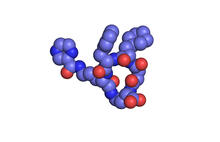
X-ray co-crystal structure of Src kinase bound to a DNA-templated macrocycle inhibitor 1
3413
X-ray co-crystal structure of Src kinase bound to a DNA-templated macrocycle inhibitor. Markus A. Seeliger, Stony Brook University Medical School and David R. Liu, Harvard University View Media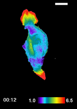
RAC1 activation in motile fibroblast
2457
Novel biosensor system maps the timing and location of Rac protein activation in a living mouse embryo fibroblast. Klaus Hahn, University of North Carolina, Chapel Hill Medical School View Media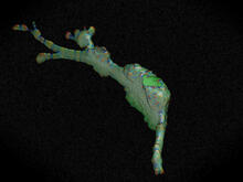
Cell curvature
2803
Rendering of the surface of an endothelial cell; membrane curvature is color coded. This is an example of NIH-supported research on single-cell analysis. Gaudenz Danuser, Harvard Medical School View Media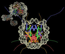
Nucleosome
2741
Like a strand of white pearls, DNA wraps around an assembly of special proteins called histones (colored) to form the nucleosome, a structure responsible for regulating genes and condensing DNA strand Karolin Luger, Colorado State University View Media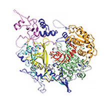
Kluyveromyces polysporus Argonaute bound to guide RNA
3408
A segment of siRNA, shown in red, guides a "slicer" protein called Argonaute (multi-colored twists and corkscrews) to the target RNA molecules. Kotaro Nakanishi and David Weinberg, Massachusetts Institute of Technology View Media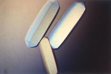
Fungal lipase (2)
2411
Crystals of fungal lipase protein created for X-ray crystallography, which can reveal detailed, three-dimensional protein structures. Alex McPherson, University of California, Irvine View Media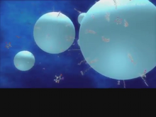
Diversity oriented synthesis: generating skeletal diversity using folding processes
3327
This 1 1/2-minute video animation was produced for chemical biologist Stuart Schreiber's lab page. The animation shows how diverse chemical structures can be produced in the lab. Eric Keller View Media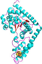
Trp_RS - tryptophanyl tRNA-synthetase family of enzymes
2483
This image represents the structure of TrpRS, a novel member of the tryptophanyl tRNA-synthetase family of enzymes. View Media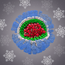
H1N1 Influenza Virus
6356
Related to image 6355. Dr. Rommie Amaro, University of California, San Diego View Media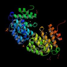
Protein from E. faecalis
2342
X-ray structure of a DNA repair enzyme superfamily representative from the human gastrointestinal bacterium Enterococcus faecalis. Midwest Center for Structural Genomics View Media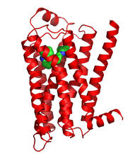
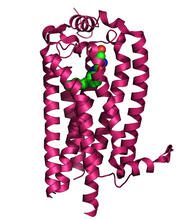
Beta 2-adrenergic receptor
3358
The receptor is shown bound to a partial inverse agonist, carazolol. Raymond Stevens, The Scripps Research Institute View Media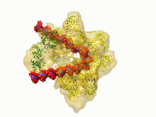
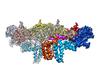
DNA replication origin recognition complex (ORC)
3307
A study published in March 2012 used cryo-electron microscopy to determine the structure of the DNA replication origin recognition complex (ORC), a semi-circular, protein complex (yellow) that recogni Huilin Li, Brookhaven National Laboratory View Media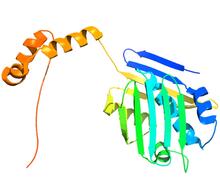
Hsp33 figure 2
3355
Featured in the March 15, 2012 issue of Biomedical Beat. Related to Hsp33 Figure 1, image 3354. Ursula Jakob and Dana Reichmann, University of Michigan View Media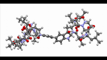
Himastatin, 360-degree view
6851
A 360-degree view of the molecule himastatin, which was first isolated from the bacterium Streptomyces himastatinicus. Himastatin shows antibiotic activity. Mohammad Movassaghi, Massachusetts Institute of Technology. View Media