Switch to Gallery View
Image and Video Gallery
This is a searchable collection of scientific photos, illustrations, and videos. The images and videos in this gallery are licensed under Creative Commons Attribution Non-Commercial ShareAlike 3.0. This license lets you remix, tweak, and build upon this work non-commercially, as long as you credit and license your new creations under identical terms.
Movie of the 19S proteasome subunit processing a protein substrate
3764
The proteasome is a critical multiprotein complex in the cell that breaks down and recycles proteins that have become damaged or are no longer needed. Andreas Martin, HHMI View Media
Diversity oriented synthesis: generating skeletal diversity using folding processes
3327
This 1 1/2-minute video animation was produced for chemical biologist Stuart Schreiber's lab page. The animation shows how diverse chemical structures can be produced in the lab. Eric Keller View Media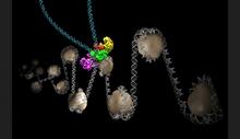
Intasome
6346
Salk researchers captured the structure of a protein complex called an intasome (center) that lets viruses similar to HIV establish permanent infection in their hosts. National Resource for Automated Molecular Microscopy http://nramm.nysbc.org/nramm-images/ Source: Bridget Carragher View MediaBeta-galactosidase montage showing cryo-EM improvement--transparent background
5882
Composite image of beta-galactosidase showing how cryo-EM’s resolution has improved dramatically in recent years. Older images to the left, more recent to the right. Veronica Falconieri, Sriram Subramaniam Lab, National Cancer Institute View Media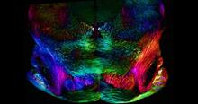
Mouse brain slice showing nerve cells
6901
A 20-µm thick section of mouse midbrain. The nerve cells are transparent and weren’t stained. Michael Shribak, Marine Biological Laboratory/University of Chicago. View Media
Sleep and the fly brain
2596
In the top snapshots, the brain of a sleep-deprived fruit fly glows orange, marking high concentrations of a synaptic protein called Bruchpilot (BRP) involved in communication between neurons. Chiara Cirelli, University of Wisconsin-Madison View Media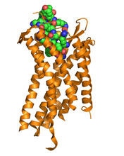
Chemokine CXCR4 receptor
3365
The receptor is shown bound to a small molecule peptide called CVX15. Raymond Stevens, The Scripps Research Institute View Media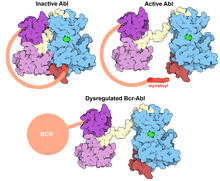
Protein kinases as cancer chemotherapy targets
7004
Protein kinases—enzymes that add phosphate groups to molecules—are cancer chemotherapy targets because they play significant roles in almost all aspects of cell function, are tightly regulated, and co Amy Wu and Christine Zardecki, RCSB Protein Data Bank. View Media
Z rings in bacterial division
2456
Lab-made liposomes contract where Z rings have gathered together and the constriction forces are greatest (arrows). Masaki Osawa, Duke University View Media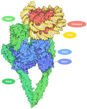
Histone deacetylases
7001
The human genome contains much of the information needed for every cell in the body to function. However, different types of cells often need different types of information. Amy Wu and Christine Zardecki, RCSB Protein Data Bank. View Media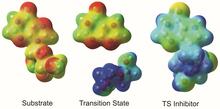
Enzyme transition states
3429
The molecule on the left is an electrostatic potential map of the van der Waals surface of the transition state for human purine nucleoside phosphorylase. Vern Schramm, Albert Einstein College of Medicine of Yeshiva University View Media
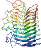
The Structure of Cilia’s Doublet Microtubules
6549
Cilia (cilium in singular) are complex molecular machines found on many of our cells. Brown Lab, Harvard Medical School and Veronica Falconieri Hays View Media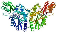
PanC from M. tuberculosis
2383
Model of an enzyme, PanC, that is involved in the last step of vitamin B5 biosynthesis in Mycobacterium tuberculosis. PanC is essential for the growth of M. Mycobacterium Tuberculosis Center, PSI View Media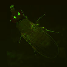
Fly by night
2417
This fruit fly expresses green fluorescent protein (GFP) in the same pattern as the period gene, a gene that regulates circadian rhythm and is expressed in all sensory neurons on the surface of the fl Jay Hirsh, University of Virginia View Media
Rabbit GPDA
2405
A crystal of rabbit GPDA protein created for X-ray crystallography, which can reveal detailed, three-dimensional protein structures. Alex McPherson, University of California, Irvine View Media
Protein crystals
1060
Structural biologists create crystals of proteins, shown here, as a first step in a process called X-ray crystallography, which can reveal detailed, three-dimensional protein structures. Alex McPherson, University of California, Irvine View Media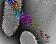
Bacterial nanowire model
6580
A model of a Geobacter sulfurreducens nanowire created from cryo-electron microscopy images. Edward Egelman, University of Virginia. View Media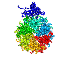
Bacteriophage P22 capsid, detail
5875
Detail of a subunit of the capsid, or outer cover, of bacteriophage P22, a virus that infects the Salmonella bacteria. Dr. Wah Chiu, Baylor College of Medicine View Media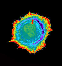
Seeing signaling protein activation in cells 01
2451
Cdc42, a member of the Rho family of small guanosine triphosphatase (GTPase) proteins, regulates multiple cell functions, including motility, proliferation, apoptosis, and cell morphology. Klaus Hahn, University of North Carolina, Chapel Hill Medical School View Media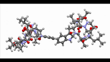
Himastatin, 360-degree view
6851
A 360-degree view of the molecule himastatin, which was first isolated from the bacterium Streptomyces himastatinicus. Himastatin shows antibiotic activity. Mohammad Movassaghi, Massachusetts Institute of Technology. View Media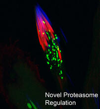
Proteasome
3451
This fruit fly spermatid recycles various molecules, including malformed or damaged proteins. Sigi Benjamin-Hong, Rockefeller University View Media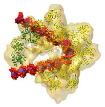
DNA replication origin recognition complex (ORC)
3597
A study published in March 2012 used cryo-electron microscopy to determine the structure of the DNA replication origin recognition complex (ORC), a semi-circular, protein complex (yellow) that recogni Huilin Li, Brookhaven National Laboratory View Media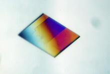
Bacterial alpha amylase
2401
A crystal of bacterial alpha amylase protein created for X-ray crystallography, which can reveal detailed, three-dimensional protein structures. Alex McPherson, University of California, Irvine View Media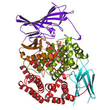
Aminopeptidase N from N. meningitidis
2341
Model of the enzyme aminopeptidase N from the human pathogen Neisseria meningitidis, which can cause meningitis epidemics. Midwest Center for Structural Genomics, PSI View Media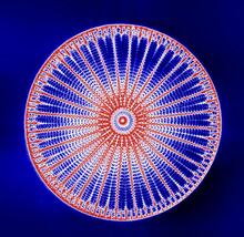
Arachnoidiscus diatom
6902
An Arachnoidiscus diatom with a diameter of 190µm. Michael Shribak, Marine Biological Laboratory/University of Chicago. View Media
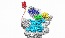
Kluyveromyces polysporus Argonaute bound to guide RNA
3408
A segment of siRNA, shown in red, guides a "slicer" protein called Argonaute (multi-colored twists and corkscrews) to the target RNA molecules. Kotaro Nakanishi and David Weinberg, Massachusetts Institute of Technology View Media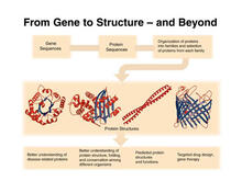
PSI: from genes to structures
2363
The goal of the Protein Structure Initiative (PSI) is to determine the three-dimensional shapes of a wide range of proteins by solving the structures of representative members of each protein family f National Institute of General Medical Sciences View MediaNuclear Lamina – Three Views
6573
Three views of the entire nuclear lamina of a HeLa cell produced by tilted light sheet 3D single-molecule super-resolution imaging using a platform termed TILT3D. Anna-Karin Gustavsson, Ph.D. View Media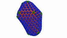
Atomic-level structure of the HIV capsid
6601
This animation shows atoms of the HIV capsid, the shell that encloses the virus's genetic material. Juan R. Perilla and the Theoretical and Computational Biophysics Group, University of Illinois at Urbana-Champaign View Media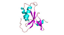
Antitoxin GhoS (Illustration 1)
3427
Structure of the bacterial antitoxin protein GhoS. GhoS inhibits the production of a bacterial toxin, GhoT, which can contribute to antibiotic resistance. Rebecca Page and Wolfgang Peti, Brown University and Thomas K. Wood, Pennsylvania State University View Media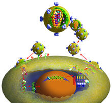
Life of an AIDS virus
2513
HIV is a retrovirus, a type of virus that carries its genetic material not as DNA but as RNA. Crabtree + Company View Media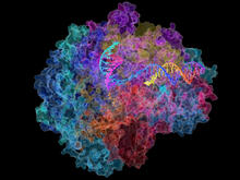
RNA Polymerase II
2484
NIGMS-funded researchers led by Roger Kornberg solved the structure of RNA polymerase II. David Bushnell, Ken Westover and Roger Kornberg, Stanford University View Media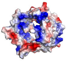
VDAC-1 (3)
2494
The structure of the pore-forming protein VDAC-1 from humans. Gerhard Wagner, Harvard Medical School View Media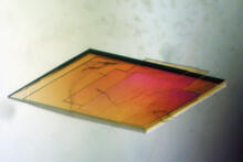
Pig trypsin crystal
2403
A crystal of pig trypsin protein created for X-ray crystallography, which can reveal detailed, three-dimensional protein structures. Alex McPherson, University of California, Irvine View Media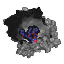
X-ray co-crystal structure of Src kinase bound to a DNA-templated macrocycle inhibitor 3
3415
X-ray co-crystal structure of Src kinase bound to a DNA-templated macrocycle inhibitor. Markus A. Seeliger, Stony Brook University Medical School and David R. Liu, Harvard University View Media
Partial Model of a Cilium’s Doublet Microtubule
6548
Cilia (cilium in singular) are complex molecular machines found on many of our cells. Brown Lab, Harvard Medical School and Veronica Falconieri Hays. View Media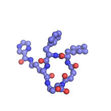
X-ray co-crystal structure of Src kinase bound to a DNA-templated macrocycle inhibitor 6
3418
X-ray co-crystal structure of Src kinase bound to a DNA-templated macrocycle inhibitor. Markus A. Seeliger, Stony Brook University Medical School and David R. Liu, Harvard University View Media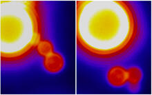
Dynamin Fission
3448
Time lapse series shows short dynamin assemblies (not visible) constricting a lipid tube to make a "beads on a string" appearance, then cutting off one of the beads i.e., catalyzing membrane fission). Ramachandran, Pucadyil et al. , The Scripps Research Institute View Media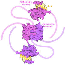
SARS-CoV-2 nucleocapsid dimer
6991
In SARS-CoV-2, the virus that causes COVID-19, nucleocapsid is a complex molecule with many functional parts. Amy Wu and Christine Zardecki, RCSB Protein Data Bank. View Media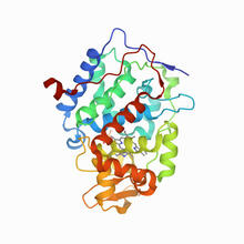
CCP enzyme
6762
The enzyme CCP is found in the mitochondria of baker’s yeast. Scientists study the chemical reactions that CCP triggers, which involve a water molecule, iron, and oxygen. Protein Data Bank. View Media
Trp_RS - tryptophanyl tRNA-synthetase family of enzymes
2483
This image represents the structure of TrpRS, a novel member of the tryptophanyl tRNA-synthetase family of enzymes. View Media
Zinc finger
2426
The structure of a gene-regulating zinc finger protein bound to DNA. Jeremy M. Berg, National Institute of General Medical Sciences View Media
Structure of amyloid-forming prion protein
3542
This structure from an amyloid-forming prion protein shows one way beta sheets can stack. Douglas Fowler, University of Washington View Media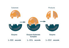
Enzymes convert subtrates into products (with labels)
2522
Enzymes convert substrates into products very quickly. See image 2521 for an unlabeled version of this illustration. Featured in The Chemistry of Health. Crabtree + Company View Media
Transient receptor potential channel TRPV5
6577
A 3D reconstruction of a transient receptor potential channel called TRPV5 that was created based on cryo-electron microscopy images. Vera Moiseenkova-Bell, University of Pennsylvania. View Media
Early life of a protein
2740
This illustration represents the early life of a protein—specifically, apomyoglobin—as it is synthesized by a ribosome and emerges from the ribosomal tunnel, which contains the newly formed protein's Silvia Cavagnero, University of Wisconsin, Madison View Media
Ribbon diagram of a cefotaxime-CCD-1 complex
6766
CCD-1 is an enzyme produced by the bacterium Clostridioides difficile that helps it resist antibiotics. Keith Hodgson, Stanford University. View MediaNuclear Lamina
6572
The 3D single-molecule super-resolution reconstruction of the entire nuclear lamina in a HeLa cell was acquired using the TILT3D platform. Anna-Karin Gustavsson, Ph.D. View Media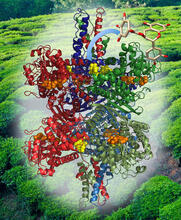
Structure of Glutamate Dehydrogenase
3421
Some children are born with a mutation in a regulatory site on this enzyme that causes them to over-secrete insulin when they consume protein. Judy Coyle, Donald Danforth Plant Science Center View Media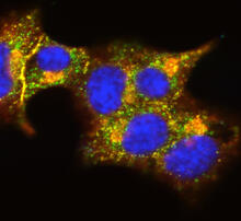
Insulin and protein interact in pancreatic beta cells
3546
A large number of proteins interact with the hormone insulin as it is produced in and secreted from the beta cells of the pancreas. William E. Balch, The Scripps Research Institute View Media