Switch to Gallery View
Image and Video Gallery
This is a searchable collection of scientific photos, illustrations, and videos. The images and videos in this gallery are licensed under Creative Commons Attribution Non-Commercial ShareAlike 3.0. This license lets you remix, tweak, and build upon this work non-commercially, as long as you credit and license your new creations under identical terms.
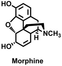
Morphine Structure
3438
The chemical structure of the morphine molecule Judy Coyle, Donald Danforth Plant Science Center View Media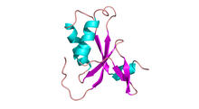
Antitoxin GhoS (Illustration 1)
3427
Structure of the bacterial antitoxin protein GhoS. GhoS inhibits the production of a bacterial toxin, GhoT, which can contribute to antibiotic resistance. Rebecca Page and Wolfgang Peti, Brown University and Thomas K. Wood, Pennsylvania State University View Media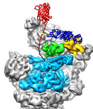
The 26S proteasome engages with a protein substrate
3763
The proteasome is a critical multiprotein complex in the cell that breaks down and recycles proteins that have become damaged or are no longer needed. Andreas Martin, HHMI View Media
PanB from M. tuberculosis (1)
2380
Model of an enzyme, PanB, from Mycobacterium tuberculosis, the bacterium that causes most cases of tuberculosis. This enzyme is an attractive drug target. Mycobacterium Tuberculosis Center, PSI View Media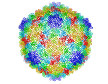
Bacteriophage P22 capsid
5874
Cryo-electron microscopy (cryo-EM) has the power to capture details of proteins and other small biological structures at the molecular level. This image shows proteins in the capsid, or outer co Dr. Wah Chiu, Baylor College of Medicine View Media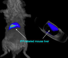
Mouse liver labeled with fluorescent probe
2601
A mouse liver glows after being tagged with specially designed infrared-fluorescent protein (IFP). Xiaokun Shu, University of California, San Diego View Media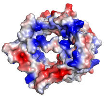
VDAC-1 (2)
2491
The structure of the pore-forming protein VDAC-1 from humans. Gerhard Wagner, Harvard Medical School View Media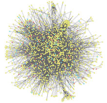
Molecular interactions
2743
This network map shows molecular interactions (yellow) associated with a congenital condition that causes heart arrhythmias and the targets for drugs that alter these interactions (red and blue). Ravi Iyengar, Mount Sinai School of Medicine View Media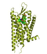
Kappa opioid receptor
3359
The receptor is shown bound to an antagonist, JDTic. Raymond Stevens, The Scripps Research Institute View Media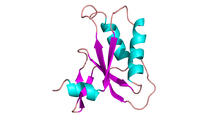
Antitoxin GhoS (Illustration 2)
3428
Structure of the bacterial antitoxin protein GhoS. GhoS inhibits the production of a bacterial toxin, GhoT, which can contribute to antibiotic resistance. Rebecca Page and Wolfgang Peti, Brown University and Thomas K. Wood, Pennsylvania State University View Media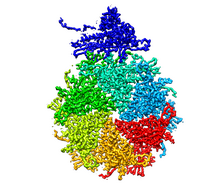
Bacteriophage P22 capsid, detail
5875
Detail of a subunit of the capsid, or outer cover, of bacteriophage P22, a virus that infects the Salmonella bacteria. Dr. Wah Chiu, Baylor College of Medicine View Media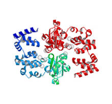
Magnesium transporter protein from E. faecalis
2345
Structure of a magnesium transporter protein from an antibiotic-resistant bacterium (Enterococcus faecalis) found in the human gut. New York Structural GenomiX Consortium View Media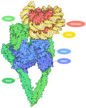
Histone deacetylases
7001
The human genome contains much of the information needed for every cell in the body to function. However, different types of cells often need different types of information. Amy Wu and Christine Zardecki, RCSB Protein Data Bank. View Media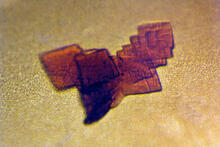
Sheep hemoglobin crystal
2392
A crystal of sheep hemoglobin protein created for X-ray crystallography, which can reveal detailed, three-dimensional protein structures. Alex McPherson, University of California, Irvine View Media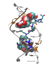
Myotonic dystrophy type 2 genetic defect
3573
Scientists revealed a detailed image of the genetic defect that causes myotonic dystrophy type 2 and used that information to design drug candidates to counteract the disease. Matthew Disney, Scripps Research Institute and Ilyas Yildirim, Northwestern University View Media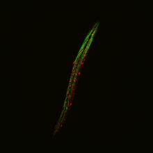
Fluorescent C. elegans showing muscle and ribosomal protein
6581
C. elegans, a tiny roundworm, with a ribosomal protein glowing red and muscle fibers glowing green. Researchers used these worms to study a molecular pathway that affects aging. Jarod Rollins, Mount Desert Island Biological Laboratory. View Media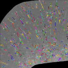
Trajectories of labeled cell receptors
2801
Trajectories of single molecule labeled cell surface receptors. This is an example of NIH-supported research on single-cell analysis. Gaudenz Danuser, Harvard Medical School View Media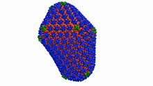
Atomic-level structure of the HIV capsid
6601
This animation shows atoms of the HIV capsid, the shell that encloses the virus's genetic material. Juan R. Perilla and the Theoretical and Computational Biophysics Group, University of Illinois at Urbana-Champaign View Media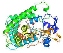
Cytochrome structure with anticancer drug
3326
This image shows the structure of the CYP17A1 enzyme (ribbons colored from blue N-terminus to red C-terminus), with the associated heme colored black. Emily Scott, University of Kansas View Media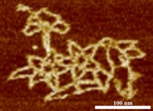
Microscopy image of bird-and-flower DNA origami
3690
An atomic force microscopy image shows DNA folded into an intricate, computer-designed structure. Hao Yan, Arizona State University View Media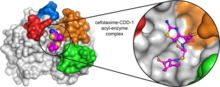
Space-filling model of a cefotaxime-CCD-1 complex
6767
CCD-1 is an enzyme produced by the bacterium Clostridioides difficile that helps it resist antibiotics. Keith Hodgson, Stanford University. View Media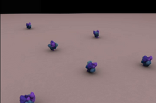
Clathrin-mediated endocytosis
5753
Endocytosis is the process by which cells are able to take up membrane and extracellular materials through the formation of a small intracellular bubble, called a vesicle. Janet Iwasa, University of Utah View Media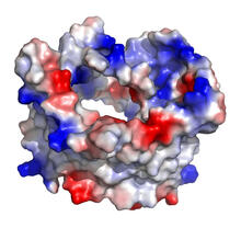
VDAC-1 (1)
2488
The structure of the pore-forming protein VDAC-1 from humans. Gerhard Wagner, Harvard Medical School View Media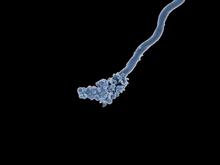
Relapsing fever bacterium (gray) and red blood cells
3585
Relapsing fever is caused by a bacterium and transmitted by certain soft-bodied ticks or body lice. The disease is seldom fatal in humans, but it can be very serious and prolonged. NIAID View Media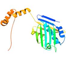
Hsp33 figure 2
3355
Featured in the March 15, 2012 issue of Biomedical Beat. Related to Hsp33 Figure 1, image 3354. Ursula Jakob and Dana Reichmann, University of Michigan View Media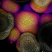
Flower-forming cells in a small plant related to cabbage (Arabidopsis)
3606
In plants, as in animals, stem cells can transform into a variety of different cell types. The stem cells at the growing tip of this Arabidopsis plant will soon become flowers. Arun Sampathkumar and Elliot Meyerowitz, California Institute of Technology View Media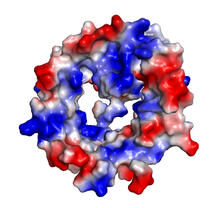
VDAC-1 (4)
2495
The structure of the pore-forming protein VDAC-1 from humans. Gerhard Wagner, Harvard Medical School View Media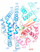
Oligoendopeptidase F from B. stearothermophilus
2373
Crystal structure of oligoendopeptidase F, a protein slicing enzyme from Bacillus stearothermophilus, a bacterium that can cause food products to spoil. Accelerated Technologies Center for Gene to 3D Structure/Midwest Center for Structural Genomics View Media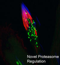
Proteasome
3451
This fruit fly spermatid recycles various molecules, including malformed or damaged proteins. Sigi Benjamin-Hong, Rockefeller University View Media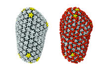
Cryo-EM reveals how the HIV capsid attaches to a human protein to evade immune detection
3755
The illustration shows the capsid of human immunodeficiency virus (HIV) whose molecular features were resolved with cryo-electron microscopy (cryo-EM). Juan R. Perilla, University of Illinois at Urbana-Champaign View Media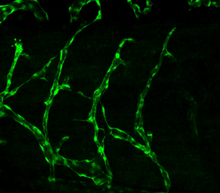
Disrupted vascular development in frog embryos
3403
Disassembly of vasculature in kdr:GFP frogs following addition of 250 µM TBZ. Related to images 3404 and 3505. Hye Ji Cha, University of Texas at Austin View Media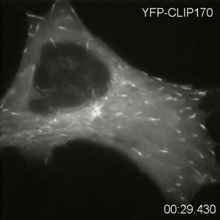
Microtubule dynamics in real time
2784
Cytoplasmic linker protein (CLIP)-170 is a microtubule plus-end-tracking protein that regulates microtubule dynamics and links microtubule ends to different intracellular structures. Gary Borisy, Marine Biology Laboratory View Media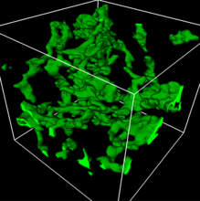
3D reconstruction of a tubular matrix in peripheral endoplasmic reticulum
5857
Detailed three-dimensional reconstruction of a tubular matrix in a thin section of the peripheral endoplasmic reticulum between the plasma membranes of the cell. Jennifer Lippincott-Schwartz, Howard Hughes Medical Institute Janelia Research Campus, Virginia View Media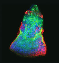
Isolated Planarian Pharynx
3593
The feeding tube, or pharynx, of a planarian worm with cilia shown in red and muscle fibers shown in green View Media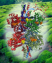
Structure of Glutamate Dehydrogenase
3421
Some children are born with a mutation in a regulatory site on this enzyme that causes them to over-secrete insulin when they consume protein. Judy Coyle, Donald Danforth Plant Science Center View Media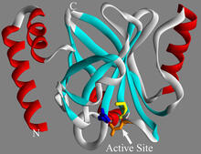
Sortase b from B. anthracis
2386
Structure of sortase b from the bacterium B. anthracis, which causes anthrax. Sortase b is an enzyme used to rob red blood cells of iron, which the bacteria need to survive. Midwest Center for Structural Genomics, PSI View Media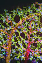
Weblike sheath covering developing egg chambers in a giant grasshopper
3616
The lubber grasshopper, found throughout the southern United States, is frequently used in biology classes to teach students about the respiratory system of insects. Kevin Edwards, Johny Shajahan, and Doug Whitman, Illinois State University. View Media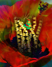
Human opioid receptor structure superimposed on poppy
3314
Opioid receptors on the surfaces of brain cells are involved in pleasure, pain, addiction, depression, psychosis, and other conditions. Raymond Stevens, The Scripps Research Institute View Media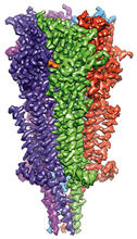
Full-length serotonin receptor (ion channel)
6579
A 3D reconstruction, created using cryo-electron microscopy, of an ion channel known as the full-length serotonin receptor in complex with the antinausea drug granisetron (orange). Sudha Chakrapani, Case Western Reserve University School of Medicine. View Media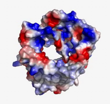
VDAC video 02
2571
This video shows the structure of the pore-forming protein VDAC-1 from humans. Gerhard Wagner, Harvard Medical School View Media
800 MHz NMR magnet
3526
Scientists use nuclear magnetic spectroscopy (NMR) to determine the detailed, 3D structures of molecules. Asokan Anbanandam, University of Kansas View Media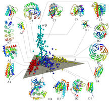
Map of protein structures 02
2367
A global "map of the protein structure universe" indicating the positions of specific proteins. Berkeley Structural Genomics Center, PSI View Media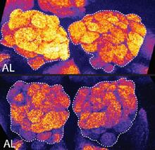
Brains of sleep-deprived and well-rested fruit flies
3490
On top, the brain of a sleep-deprived fly glows orange because of Bruchpilot, a communication protein between brain cells. These bright orange brain areas are associated with learning. Chiara Cirelli, University of Wisconsin-Madison View Media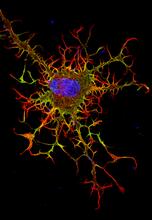
Abnormal, spiky fibroblast
3613
This is a fibroblast, a connective tissue cell that plays an important role in wound healing. Normal fibroblasts have smooth edges. Praveen Suraneni, Stowers Institute for Medical Research, Kansas City, Mo. View Media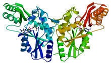
PanC from M. tuberculosis
2383
Model of an enzyme, PanC, that is involved in the last step of vitamin B5 biosynthesis in Mycobacterium tuberculosis. PanC is essential for the growth of M. Mycobacterium Tuberculosis Center, PSI View Media
Z rings in bacterial division
2456
Lab-made liposomes contract where Z rings have gathered together and the constriction forces are greatest (arrows). Masaki Osawa, Duke University View Media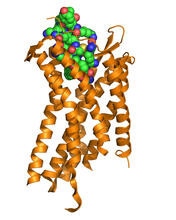
Chemokine CXCR4 receptor
3365
The receptor is shown bound to a small molecule peptide called CVX15. Raymond Stevens, The Scripps Research Institute View Media
Bacterial glucose isomerase
2409
A crystal of bacterial glucose isomerase protein created for X-ray crystallography, which can reveal detailed, three-dimensional protein structures. Alex McPherson, University of California, Irvine View Media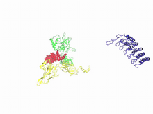
A molecular switch strips transcription factor from DNA
3729
In this video, Rice University scientists used molecular modeling with a mathematical algorithm called AWSEM (for associative memory, water-mediated, structure and energy model) and structural data to Davit Potoyan and Peter Wolynes View Media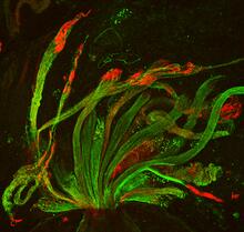
Fruit fly spermatids
3590
Developing spermatids (precursors of mature sperm cells) begin as small, round cells and mature into long-tailed, tadpole-shaped ones. Lacramioara Fabian, The Hospital for Sick Children, Toronto, Canada View Media