Switch to Gallery View
Image and Video Gallery
This is a searchable collection of scientific photos, illustrations, and videos. The images and videos in this gallery are licensed under Creative Commons Attribution Non-Commercial ShareAlike 3.0. This license lets you remix, tweak, and build upon this work non-commercially, as long as you credit and license your new creations under identical terms.
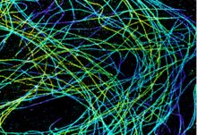
Microtubules in African green monkey cells
6891
Microtubules in African green monkey cells. Microtubules are strong, hollow fibers that provide cells with structural support. Melike Lakadamyali, Perelman School of Medicine at the University of Pennsylvania. View Media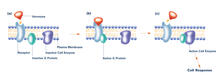
G switch (with labels and stages)
2538
The G switch allows our bodies to respond rapidly to hormones. G proteins act like relay batons to pass messages from circulating hormones into cells. Crabtree + Company View Media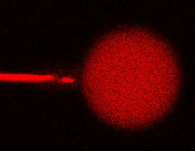
Bee venom toxin destroying a cell
3583
This video condenses 6.5 minutes into less than a minute to show how the toxin in bee venom, called melittin, destroys an animal or bacterial cell. Huey Huang, Rice University View Media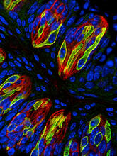
Taste buds signal different tastes through ATP release
3444
Taste buds in a mouse tongue epithelium with types I, II, and III taste cells visualized by cell-type-specific fluorescent antibodies. Aki Taruno, Perelman School of Medicine, University of Pennsylvania View Media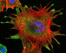
mDia1 antibody staining- 02
3331
Cells move forward with lamellipodia and filopodia supported by networks and bundles of actin filaments. Proper, controlled cell movement is a complex process. Rong Li and Praveen Suraneni, Stowers Institute for Medical Research View Media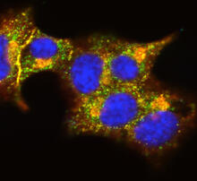
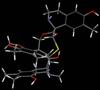
Insulin and protein interact in pancreatic beta cells
3546
A large number of proteins interact with the hormone insulin as it is produced in and secreted from the beta cells of the pancreas. William E. Balch, The Scripps Research Institute View Media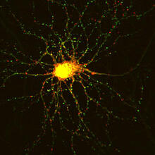
Neuron with labeled synapses
3509
In this image, recombinant probes known as FingRs (Fibronectin Intrabodies Generated by mRNA display) were expressed in a cortical neuron, where they attached fluorescent proteins to either PSD95 (gre Don Arnold and Richard Roberts, University of Southern California. View Media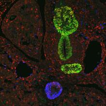
Transcription factor Sox17 controls embryonic development of certain internal organs
3440
During embryonic development, transcription factors (proteins that regulate gene expression) govern the differentiation of cells into separate tissues and organs. James M. Wells, Cincinnati Children's Hospital Medical Center View Media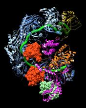
CRISPR surveillance complex
6352
This image shows how the CRISPR surveillance complex is disabled by two copies of anti-CRISPR protein AcrF1 (red) and one AcrF2 (light green). NRAMM National Resource for Automated Molecular Microscopy http://nramm.nysbc.org/nramm-images/ Source: Bridget Carragher View Media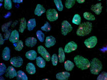
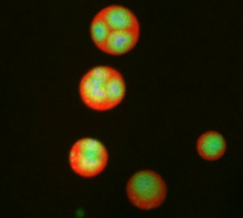
Induced pluripotent stem cells from skin 02
3279
These induced pluripotent stem cells (iPS cells) were derived from a woman's skin. Blue show nuclei. Green show a protein found in iPS cells but not in skin cells (NANOG). Kathrin Plath lab, University of California, Los Angeles, via CIRM View Media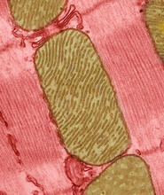
Mitochondria from rat heart muscle cell_2
3664
These mitochondria (brown) are from the heart muscle cell of a rat. Mitochondria have an inner membrane that folds in many places (and that appears here as striations). National Center for Microscopy and Imaging Research View Media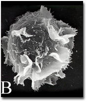
Activated mast cell surface
2637
A scanning electron microscope image of an activated mast cell. This image illustrates the interesting topography of the cell membrane, which is populated with receptors. Bridget Wilson, University of New Mexico View Media
Telomerase illustration
1335
Reactivating telomerase in our cells does not appear to be a good way to extend the human lifespan. Cancer cells reactivate telomerase. Judith Stoffer View Media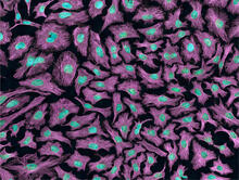
HeLa cells
3520
Multiphoton fluorescence image of HeLa cells with cytoskeletal microtubules (magenta) and DNA (cyan). Nikon RTS2000MP custom laser scanning microscope. National Center for Microscopy and Imaging Research (NCMIR) View Media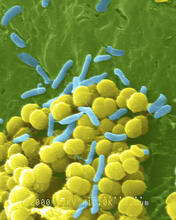
Bacteria shapes
1158
A colorized scanning electron micrograph of bacteria. Scanning electron microscopes allow scientists to see the three-dimensional surface of their samples. Tina Weatherby Carvalho, University of Hawaii at Manoa View Media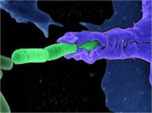
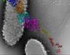
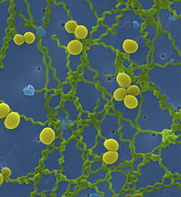
Anthrax bacteria (green) being swallowed by an immune system cell
3612
Multiple anthrax bacteria (green) being enveloped by an immune system cell (purple). Anthrax bacteria live in soil and form dormant spores that can survive for decades. Camenzind G. Robinson, Sarah Guilman, and Arthur Friedlander, United States Army Medical Research Institute of Infectious Diseases View Media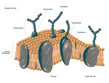
Plasma membrane (with labels)
2524
The plasma membrane is a cell's protective barrier. See image 2523 for an unlabeled version of this illustration. Featured in The Chemistry of Health. Crabtree + Company View Media
Leptospira bacteria
1166
Leptospira, shown here in green, is a type (genus) of elongated, spiral-shaped bacteria. Infection can cause Weil's disease, a kind of jaundice, in humans. Tina Weatherby Carvalho, University of Hawaii at Manoa View Media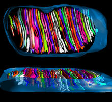
Mitochondrion from insect flight muscle
3662
This is a tomographic reconstruction of a mitochondrion from an insect flight muscle. National Center for Microscopy and Imaging Research View Media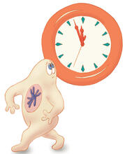
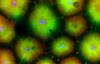
Nucleolus subcompartments spontaneously self-assemble 3
3792
What looks a little like distant planets with some mysterious surface features are actually assemblies of proteins normally found in the cell's nucleolus, a small but very important protein complex lo Nilesh Vaidya, Princeton University View Media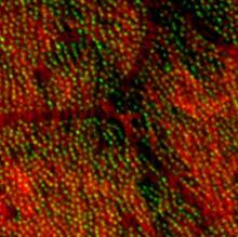
Centrioles anchor cilia in planaria
3292
Centrioles (green) anchor cilia (red), which project on the surface of pharynx cells of the freshwater planarian Schmidtea mediterranea. Juliette Azimzadeh, University of California, San Francisco View Media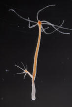
Hydra 03
2439
Hydra magnipapillata is an invertebrate animal used as a model organism to study developmental questions, for example the formation of the body axis. Hiroshi Shimizu, National Institute of Genetics in Mishima, Japan View Media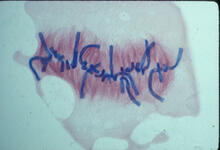
Lily mitosis 08
1021
A light microscope image of a cell from the endosperm of an African globe lily (Scadoxus katherinae). This is one frame of a time-lapse sequence that shows cell division in action. Andrew S. Bajer, University of Oregon, Eugene View Media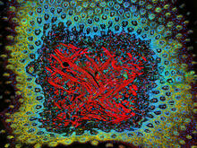
Tongue 1
5810
Microscopy image of tongue. One in a series of two, see image 5811 National Center for Microscopy and Imaging Research (NCMIR) View Media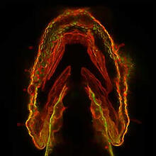
Glowing glycans
2473
Sugars light up the cells in this jaw of a 3-day-old zebrafish embryo and highlight a scientific first: labeling and tracking the movements of sugar chains called glycans in a living organism. Carolyn Bertozzi, University of California, Berkeley View Media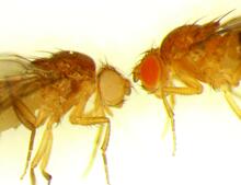
Drosophila
6344
Two adult fruit flies (Drosophila) Dr. Vicki Losick, MDI Biological Laboratory, www.mdibl.org View Media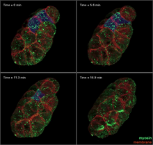
Four timepoints in gastrulation
3334
It has been said that gastrulation is the most important event in a person's life. Bob Goldstein, University of North Carolina, Chapel Hill View Media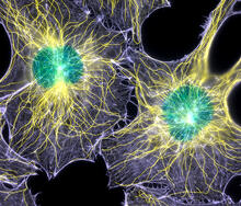
Colorful cells
2428
Actin (purple), microtubules (yellow), and nuclei (green) are labeled in these cells by immunofluorescence. This image won first place in the Nikon 2003 Small World photo competition. Torsten Wittmann, Scripps Research Institute View Media
Blinking bacteria
2724
Like a pulsing blue shower, E. coli cells flash in synchrony. Genes inserted into each cell turn a fluorescent protein on and off at regular intervals. Jeff Hasty, University of California, San Diego View Media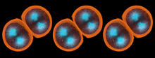
Sea urchin embryo 01
1047
Stereo triplet of a sea urchin embryo stained to reveal actin filaments (orange) and microtubules (blue). George von Dassow, University of Washington View Media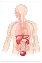
Body toxins
2496
Body organs such as the liver and kidneys process chemicals and toxins. These "target" organs are susceptible to damage caused by these substances. Crabtree + Company View Media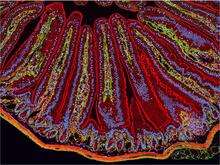
NCMIR Intestine-1
3389
The small intestine is where most of our nutrients from the food we eat are absorbed into the bloodstream. Tom Deerinck, National Center for Microscopy and Imaging Research (NCMIR) View Media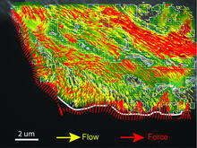
Intracellular forces
2799
Force vectors computed from actin cytoskeleton flow. This is an example of NIH-supported research on single-cell analysis. Gaudenz Danuser, Harvard Medical School View Media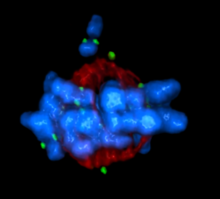
Mitotic cell awaits chromosome alignment
5765
During mitosis, spindle microtubules (red) attach to chromosome pairs (blue), directing them to the spindle equator. View Media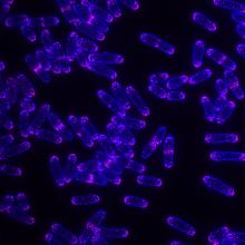
Yeast cells with Fimbrin Fim1
6794
Yeast cells with the protein Fimbrin Fim1 shown in magenta. This protein plays a role in cell division. This image was captured using wide-field microscopy with deconvolution.Alaina Willet, Kathy Gould’s lab, Vanderbilt University. View Media
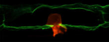
Anchor cell in basement membrane
2707
An anchor cell (red) pushes through the basement membrane (green) that surrounds it. Elliott Hagedorn, Duke University. View Media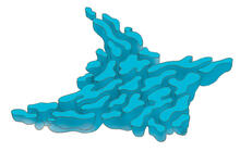
Smooth ER
1292
The endoplasmic reticulum comes in two types: Rough ER is covered with ribosomes and prepares newly made proteins; smooth ER specializes in making lipids and breaking down toxic molecules. Judith Stoffer View Media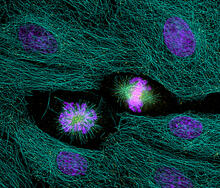
Highlighted cells
2429
The cytoskeleton (green) and DNA (purple) are highlighed in these cells by immunofluorescence. Torsten Wittmann, Scripps Research Institute View Media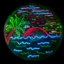
Glowing bacteria make a pretty postcard
3492
This tropical scene, reminiscent of a postcard from Key West, is actually a petri dish containing an artistic arrangement of genetically engineered bacteria. Nathan C. Shaner, The Scintillon Institute View Media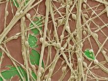
Synapses in culture
3399
Cultured hippocampal neurons grown on a substrate of glial cells (astrocytes). The glial cells form the pink/brown underlayment in this image. The tan threads are the neurons. National Center for Microscopy and Imaging Research View MediaGenetic mosaicism in fruit flies
6983
Fat tissue from the abdomen of a genetically mosaic adult fruit fly. Genetic mosaicism means that the fly has cells with different genotypes even though it formed from a single zygote. Akhila Rajan, Fred Hutchinson Cancer Center View Media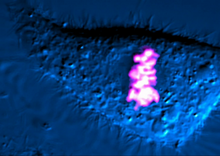
Dividing cell
6965
As this cell was undergoing cell division, it was imaged with two microscopy techniques: differential interference contrast (DIC) and confocal. The DIC view appears in blue and shows the entire cell. Dylan T. Burnette, Vanderbilt University School of Medicine. View Media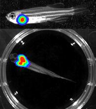
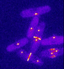
Bioluminescent imaging in adult zebrafish - lateral and overhead view
3556
Luciferase-based imaging enables visualization and quantification of internal organs and transplanted cells in live adult zebrafish. Kenneth Poss, Duke University View Media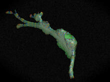
Cell curvature
2803
Rendering of the surface of an endothelial cell; membrane curvature is color coded. This is an example of NIH-supported research on single-cell analysis. Gaudenz Danuser, Harvard Medical School View Media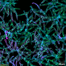
Snowflake yeast 2
6970
Multicellular yeast called snowflake yeast that researchers created through many generations of directed evolution from unicellular yeast. William Ratcliff, Georgia Institute of Technology. View Media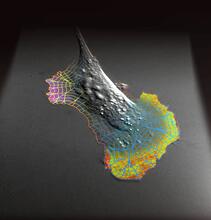
Biosensors illustration
2802
A rendering of an activity biosensor image overlaid with a cell-centered frame of reference used for image analysis of signal transduction. Gaudenz Danuser, Harvard Medical School View Media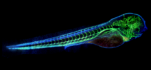
Zebrafish embryo showing vasculature
6661
A zebrafish embryo. The blue areas are cell bodies, the green lines are blood vessels, and the red glow is blood. Kevin Eliceiri, University of Wisconsin-Madison. View Media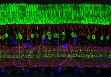
The eye uses many layers of nerve cells to convert light into sight
3635
This image captures the many layers of nerve cells in the retina. The top layer (green) is made up of cells called photoreceptors that convert light into electrical signals to relay to the brain. Wei Li, National Eye Institute, National Institutes of Health View Media