Switch to Gallery View
Image and Video Gallery
This is a searchable collection of scientific photos, illustrations, and videos. The images and videos in this gallery are licensed under Creative Commons Attribution Non-Commercial ShareAlike 3.0. This license lets you remix, tweak, and build upon this work non-commercially, as long as you credit and license your new creations under identical terms.
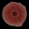
Snowflake yeast 2
6970
Multicellular yeast called snowflake yeast that researchers created through many generations of directed evolution from unicellular yeast. William Ratcliff, Georgia Institute of Technology. View Media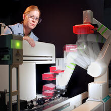
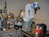
Student overseeing protein cloning robot
2356
Student Christina Hueneke of the Midwest Center for Structural Genomics is overseeing a protein cloning robot. Midwest Center for Structural Genomics View Media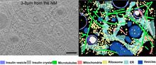
Cryo-ET cross-section of a rat pancreas cell
6608
On the left, a cross-section slice of a rat pancreas cell captured using cryo-electron tomography (cryo-ET). On the right, a 3D, color-coded version of the image highlighting cell structures. Xianjun Zhang, University of Southern California. View Media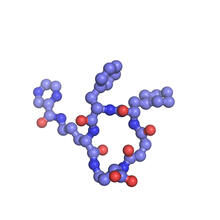
X-ray co-crystal structure of Src kinase bound to a DNA-templated macrocycle inhibitor 6
3418
X-ray co-crystal structure of Src kinase bound to a DNA-templated macrocycle inhibitor. Markus A. Seeliger, Stony Brook University Medical School and David R. Liu, Harvard University View Media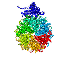
Bacteriophage P22 capsid, detail
5875
Detail of a subunit of the capsid, or outer cover, of bacteriophage P22, a virus that infects the Salmonella bacteria. Dr. Wah Chiu, Baylor College of Medicine View Media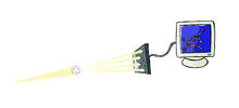
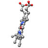
X-ray crystallography
2511
X-ray crystallography allows researchers to see structures too small to be seen by even the most powerful microscopes. Crabtree + Company View Media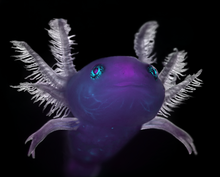
Axolotl
6932
An axolotl—a type of salamander—that has been genetically modified so that its developing nervous system glows purple and its Schwann cell nuclei appear light blue. Prayag Murawala, MDI Biological Laboratory and Hannover Medical School. View Media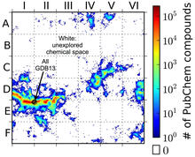
Computer algorithm
3458
This computer algorithm plots all feasible small carbon-based molecules as though they were cities on a map and identifies huge, unexplored spaces that may help fuel research into new drug therapies. Aaron Virshup, Julia Contreras-Garcia, Peter Wipf, Weitao Yang and David Beratan, University of Pittsburgh Center for Chemical Methodologies and Library Development View Media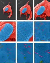
Crab larva eye
1251
Colorized scanning electron micrographs progressively zoom in on the eye of a crab larva. In the higher-resolution frames, bacteria are visible on the eye. Tina Weatherby Carvalho, University of Hawaii at Manoa View MediaNuclear Lamina – Three Views
6573
Three views of the entire nuclear lamina of a HeLa cell produced by tilted light sheet 3D single-molecule super-resolution imaging using a platform termed TILT3D. Anna-Karin Gustavsson, Ph.D. View Media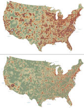
Mapping disease spread
2320
How far and fast an infectious disease spreads across a community depends on many factors, including transportation. These U.S. David Chrest, RTI International View Media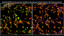
Single-cell “radios” video
7022
Individual cells are color-coded based on their identity and signaling activity using a protein circuit technology developed by the Coyle Lab. Scott Coyle, University of Wisconsin-Madison. View Media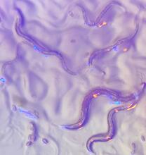
C. elegans with blue and yellow lights in the background
6750
These microscopic roundworms, called Caenorhabditis elegans, lack eyes and the opsin proteins used by visual systems to detect colors. H. Robert Horvitz and Dipon Ghosh, Massachusetts Institute of Technology. View Media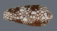
Cone snail shell
2576
A shell from the venomous cone snail Conus omaria, which lives in the Pacific and Indian oceans and eats other snails. Kerry Matz, University of Utah View Media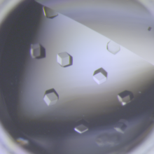
Crystals of CCD-1 in complex with cefotaxime
6764
CCD-1 is an enzyme produced by the bacterium Clostridioides difficile that helps it resist antibiotics. Keith Hodgson, Stanford University. View Media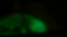
Glow-in-the-dark salamanders
2715
These six-month-old axolotls, a kind of salamander, glow green and blue under ultraviolet light. That's because they were genetically modified to make harmless green fluorescent protein, or GFP. View Media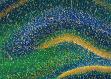
Rat Hippocampus
3308
This image of the hippocampus was taken with an ultra-widefield high-speed multiphoton laser microscope. Tom Deerinck, NCMIR View Media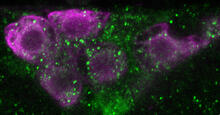
Insulin production and fat sensing in fruit flies
6982
Fourteen neurons (magenta) in the adult Drosophila brain produce insulin, and fat tissue sends packets of lipids to the brain via the lipoprotein carriers (green). Akhila Rajan, Fred Hutchinson Cancer Center View Media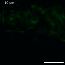
Vimentin in a quail embryo
2809
Video of high-resolution confocal images depicting vimentin immunofluorescence (green) and nuclei (blue) at the edge of a quail embryo yolk. Andrés Garcia, Georgia Tech View Media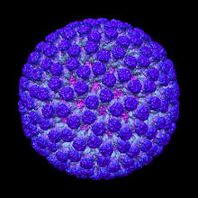
Rotavirus structure
3584
This image shows a computer-generated, three-dimensional map of the rotavirus structure. This virus infects humans and other animals and causes severe diarrhea in infants and young children. Bridget Carragher, The Scripps Research Institute, La Jolla, CA View Media
Adult Hawaiian bobtail squid burying in the sand
7012
Each morning, the nocturnal Hawaiian bobtail squid, Euprymna scolopes, hides from predators by digging into the sand. At dusk, it leaves the sand again to hunt. Margaret J. McFall-Ngai, Carnegie Institution for Science/California Institute of Technology, and Edward G. Ruby, California Institute of Technology. View Media