Switch to Gallery View
Image and Video Gallery
This is a searchable collection of scientific photos, illustrations, and videos. The images and videos in this gallery are licensed under Creative Commons Attribution Non-Commercial ShareAlike 3.0. This license lets you remix, tweak, and build upon this work non-commercially, as long as you credit and license your new creations under identical terms.
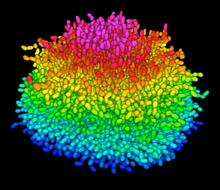
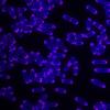
A Growing Bacterial Biofilm
5825
A growing Vibrio cholerae (cholera) biofilm. Cholera bacteria form colonies called biofilms that enable them to resist antibiotic therapy within the body and other challenges to their growth. Jing Yan, Ph.D., and Bonnie Bassler, Ph.D., Department of Molecular Biology, Princeton University, Princeton, NJ. View Media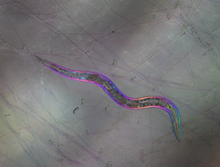
C. elegans trapped by carnivorous fungus
6963
Real-time footage of Caenorhabditis elegans, a tiny roundworm, trapped by a carnivorous fungus, Arthrobotrys dactyloides. Michael Shribak, Marine Biological Laboratory/University of Chicago. View Media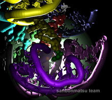
Natural nanomachine in action
2336
Using a supercomputer to simulate the movement of atoms in a ribosome, researchers looked into the core of this protein-making nanomachine and took snapshots. Kevin Sanbonmatsu, Los Alamos National Laboratory View Media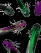
Axolotls showing nervous system components
6928
Axolotls—a type of salamander—that have been genetically modified so that various parts of their nervous systems glow purple and green. Prayag Murawala, MDI Biological Laboratory and Hannover Medical School. View MediaBeta-galactosidase montage showing cryo-EM improvement--transparent background
5882
Composite image of beta-galactosidase showing how cryo-EM’s resolution has improved dramatically in recent years. Older images to the left, more recent to the right. Veronica Falconieri, Sriram Subramaniam Lab, National Cancer Institute View Media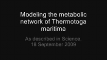
Thermotoga maritima and its metabolic network
2702
A combination of protein structures determined experimentally and computationally shows us the complete metabolic network of a heat-loving bacterium. View Media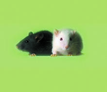
Lab mice
1069
Many researchers use the mouse (Mus musculus) as a model organism to study mammalian biology. Bill Branson, National Institutes of Health View Media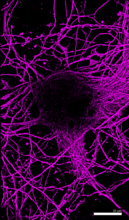
Microtubules in hippocampal neurons
6890
Microtubules (magenta) in neurons of the hippocampus, a part of the brain involved in learning and memory. Microtubules are strong, hollow fibers that provide structural support to cells. Melike Lakadamyali, Perelman School of Medicine at the University of Pennsylvania. View Media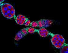
Confocal microscopy image of two Drosophila ovarioles
5772
Ovarioles in female insects are tubes in which egg cells (called oocytes) form at one end and complete their development as they reach the other end of the tube. 2004 Olympus BioScapes Competition View Media
Leading cells with light
2708
A blue laser beam turns on a protein that helps this human cancer cell move. Responding to the stimulus, the protein, called Rac1, first creates ruffles at the edge of the cell. Yi Wu, University of North Carolina View Media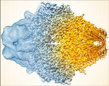
Beta-galactosidase montage showing cryo-EM improvement--gradient background
5883
Composite image of beta-galactosidase showing how cryo-EM’s resolution has improved dramatically in recent years. Older images to the left, more recent to the right. Veronica Falconieri, Sriram Subramaniam Lab, National Cancer Institute View Media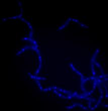
Bacillus anthracis being killed
3525
Bacillus anthracis (anthrax) cells being killed by a fluorescent trans-translation inhibitor, which disrupts bacterial protein synthesis. Kenneth Keiler, Penn State University View Media
Chromatin in human tenocyte
6893
The nucleus of a degenerating human tendon cell, also known as a tenocyte. It has been color-coded based on the density of chromatin—a substance made up of DNA and proteins. Melike Lakadamyali, Perelman School of Medicine at the University of Pennsylvania. View Media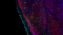
Video of Calling Cards in a mouse brain
6781
The green spots in this mouse brain are cells labeled with Calling Cards, a technology that records molecular events in brain cells as they mature. NIH Director's Blog View Media
Rabbit GPDA
2405
A crystal of rabbit GPDA protein created for X-ray crystallography, which can reveal detailed, three-dimensional protein structures. Alex McPherson, University of California, Irvine View Media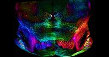
Mouse brain slice showing nerve cells
6901
A 20-µm thick section of mouse midbrain. The nerve cells are transparent and weren’t stained. Michael Shribak, Marine Biological Laboratory/University of Chicago. View Media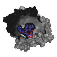
X-ray co-crystal structure of Src kinase bound to a DNA-templated macrocycle inhibitor 3
3415
X-ray co-crystal structure of Src kinase bound to a DNA-templated macrocycle inhibitor. Markus A. Seeliger, Stony Brook University Medical School and David R. Liu, Harvard University View Media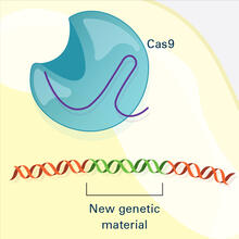
CRISPR Illustration Frame 4
6488
This illustration shows, in simplified terms, how the CRISPR-Cas9 system can be used as a gene-editing tool. National Institute of General Medical Sciences. View Media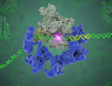
TFIID complex binds DNA to start gene transcription
3766
Gene transcription is a process by which the genetic information encoded in DNA is transcribed into RNA. Eva Nogales, Berkeley Lab View Media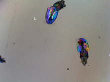
Young squids
6903
Real-time movie of young squids. Michael Shribak, Marine Biological Laboratory/University of Chicago. View Media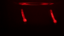
Culex quinquefasciatus mosquito larvae
6771
Mosquito larvae with genes edited by CRISPR swimming in water. Valentino Gantz, University of California, San Diego. View Media
Breast cancer cells change migration phenotypes
6986
Cancer cells can change their migration phenotype, which includes their shape and the way that they move to invade different tissues. Bo Sun, Oregon State University. View Media
Microarray 01
1070
Microarrays, also called gene chips, are tools that let scientists track the activity of hundreds or thousands of genes simultaneously. Maggie Werner-Washburne, University of New Mexico, Albuquerque View Media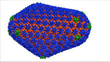
HIV Capsid
3477
This image is a computer-generated model of the approximately 4.2 million atoms of the HIV capsid, the shell that contains the virus' genetic material. Juan R. Perilla and the Theoretical and Computational Biophysics Group, University of Illinois at Urbana-Champaign View Media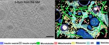
Cryo-ET cross-section of a rat pancreas cell
6608
On the left, a cross-section slice of a rat pancreas cell captured using cryo-electron tomography (cryo-ET). On the right, a 3D, color-coded version of the image highlighting cell structures. Xianjun Zhang, University of Southern California. View Media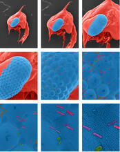
Crab larva eye
1251
Colorized scanning electron micrographs progressively zoom in on the eye of a crab larva. In the higher-resolution frames, bacteria are visible on the eye. Tina Weatherby Carvalho, University of Hawaii at Manoa View Media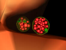
Multivesicular bodies containing intralumenal vesicles assemble at the vacuole 3
5767
Collecting and transporting cellular waste and sorting it into recylable and nonrecylable pieces is a complex business in the cell. Matthew West and Greg Odorizzi, University of Colorado View Media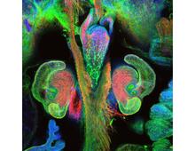
The nascent juvenile light organ of the Hawaiian bobtail squid
7017
A light organ (~0.5 mm across) of a Hawaiian bobtail squid, Euprymna scolopes, with different tissues are stained various colors. Margaret J. McFall-Ngai, Carnegie Institution for Science/California Institute of Technology, and Edward G. Ruby, California Institute of Technology. View Media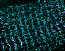
Single-Molecule Imaging
3339
This is a super-resolution light microscope image taken by Hiro Hakozaki and Masa Hoshijima of NCMIR. Tom Deerinck, NCMIR View Media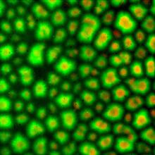
Cell-like compartments from frog eggs 3
6586
Cell-like compartments that spontaneously emerged from scrambled frog eggs. Endoplasmic reticulum (red) and microtubules (green) are visible. Image created using epifluorescence microscopy. Xianrui Cheng, Stanford University School of Medicine. View Media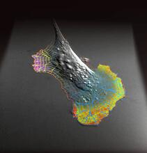
Biosensors illustration
2802
A rendering of an activity biosensor image overlaid with a cell-centered frame of reference used for image analysis of signal transduction. Gaudenz Danuser, Harvard Medical School View Media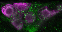
Insulin production and fat sensing in fruit flies
6982
Fourteen neurons (magenta) in the adult Drosophila brain produce insulin, and fat tissue sends packets of lipids to the brain via the lipoprotein carriers (green). Akhila Rajan, Fred Hutchinson Cancer Center View Media
Protein crystals
1060
Structural biologists create crystals of proteins, shown here, as a first step in a process called X-ray crystallography, which can reveal detailed, three-dimensional protein structures. Alex McPherson, University of California, Irvine View Media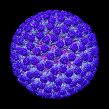
Rotavirus structure
3584
This image shows a computer-generated, three-dimensional map of the rotavirus structure. This virus infects humans and other animals and causes severe diarrhea in infants and young children. Bridget Carragher, The Scripps Research Institute, La Jolla, CA View Media
Crane fly spermatocyte undergoing meiosis
6898
A crane fly spermatocyte during metaphase of meiosis-I, a step in the production of sperm. Michael Shribak, Marine Biological Laboratory/University of Chicago. View Media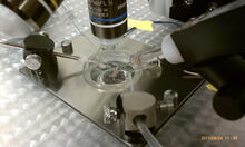
Electrode probe on mouse Huntington's muscle cell
3479
Using an electrode, researchers apply an electrical pulse onto a piece of muscle tissue affected by Huntington's disease. Grigor Varuzhanyan and Andrew A. Voss, California State Polytechnic University View Media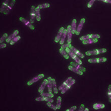
Yeast cells with endocytic actin patches
6793
Yeast cells with endocytic actin patches (green). These patches help cells take in outside material. When a cell is in interphase, patches concentrate at its ends. Alaina Willet, Kathy Gould’s lab, Vanderbilt University. View Media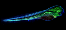
Zebrafish embryo showing vasculature
6661
A zebrafish embryo. The blue areas are cell bodies, the green lines are blood vessels, and the red glow is blood. Kevin Eliceiri, University of Wisconsin-Madison. View Media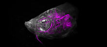
Zebrafish head vasculature video
6933
Various views of a zebrafish head with blood vessels shown in purple. Prayag Murawala, MDI Biological Laboratory and Hannover Medical School. View Media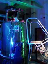
Superconducting magnet
1120
Superconducting magnet for NMR research, from the February 2003 profile of Dorothee Kern in Findings. Mike Lovett View Media
Glow-in-the-dark salamanders
2715
These six-month-old axolotls, a kind of salamander, glow green and blue under ultraviolet light. That's because they were genetically modified to make harmless green fluorescent protein, or GFP. View Media
RNase A (1)
2398
A crystal of RNase A protein created for X-ray crystallography, which can reveal detailed, three-dimensional protein structures. Alex McPherson, University of California, Irvine View Media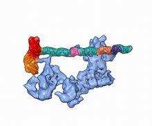
Dynamic cryo-EM model of the human transcription preinitiation complex
5730
Gene transcription is a process by which information encoded in DNA is transcribed into RNA. Eva Nogales, Berkeley Lab View Media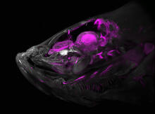
Zebrafish head vasculature
6934
A zebrafish head with blood vessels shown in purple. Prayag Murawala, MDI Biological Laboratory and Hannover Medical School. View Media
Bovine milk alpha-lactalbumin (2)
2404
Crystals of bovine milk alpha-lactalbumin protein created for X-ray crystallography, which can reveal detailed, three-dimensional protein structures. Alex McPherson, University of California, Irvine View Media
Podocytes from a chronically diseased kidney
3565
This scanning electron microscope (SEM) image shows podocytes--cells in the kidney that play a vital role in filtering waste from the bloodstream--from a patient with chronic kidney disease. Olga Troyanskaya, Princeton University and Matthias Kretzler, University of Michigan View Media
X-ray crystallography
2511
X-ray crystallography allows researchers to see structures too small to be seen by even the most powerful microscopes. Crabtree + Company View Media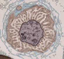
Transmission electron microscopy showing cross-section of the node of Ranvier
3740
Nodes of Ranvier are short gaps in the myelin sheath surrounding myelinated nerve cells (axons). Tom Deerinck, National Center for Microscopy and Imaging Research (NCMIR) View Media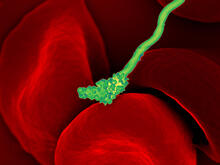
Human blood cells with Borrelia hermsii, a bacterium that causes relapsing fever
3586
Relapsing fever is caused by a bacterium and transmitted by certain soft-bodied ticks or body lice. The disease is seldom fatal in humans, but it can be very serious and prolonged. NIAID View Media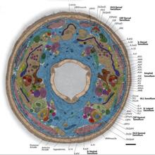
Annotated TEM cross-section of C. elegans (roundworm)
5760
The worm Caenorhabditis elegans is a popular laboratory animal because its small size and fairly simple body make it easy to study. Piali Sengupta, Brandeis University View Media