Switch to Gallery View
Image and Video Gallery
This is a searchable collection of scientific photos, illustrations, and videos. The images and videos in this gallery are licensed under Creative Commons Attribution Non-Commercial ShareAlike 3.0. This license lets you remix, tweak, and build upon this work non-commercially, as long as you credit and license your new creations under identical terms.
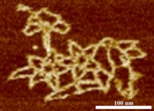
Microscopy image of bird-and-flower DNA origami
3690
An atomic force microscopy image shows DNA folded into an intricate, computer-designed structure. Hao Yan, Arizona State University View Media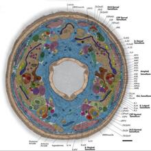
Annotated TEM cross-section of C. elegans (roundworm)
5760
The worm Caenorhabditis elegans is a popular laboratory animal because its small size and fairly simple body make it easy to study. Piali Sengupta, Brandeis University View Media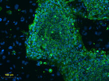
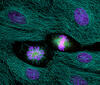
Human embryonic stem cells on feeder cells
3274
This fluorescent microscope image shows human embryonic stem cells whose nuclei are stained green. Blue staining shows the surrounding supportive feeder cells. Michael Longaker lab, Stanford University School of Medicine, via CIRM View Media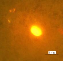
Finding one bug
2314
A nanometer-sized biosensor can detect a single deadly bacterium in tainted ground beef. How? Weihong Tan, University of Florida in Gainesville View Media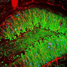
Brain showing hallmarks of Alzheimer's disease
3604
Along with blood vessels (red) and nerve cells (green), this mouse brain shows abnormal protein clumps known as plaques (blue). Alvin Gogineni, Genentech View Media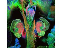
The nascent juvenile light organ of the Hawaiian bobtail squid
7017
A light organ (~0.5 mm across) of a Hawaiian bobtail squid, Euprymna scolopes, with different tissues are stained various colors. Margaret J. McFall-Ngai, Carnegie Institution for Science/California Institute of Technology, and Edward G. Ruby, California Institute of Technology. View Media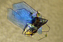
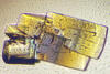
DNase
2410
Crystals of DNase protein created for X-ray crystallography, which can reveal detailed, three-dimensional protein structures. Alex McPherson, University of California, Irvine View Media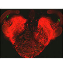
Bacterial cells aggregating above the light organ of the Hawaiian bobtail squid
7018
A light organ (~0.5 mm across) of a juvenile Hawaiian bobtail squid, Euprymna scolopes. Margaret J. McFall-Ngai, Carnegie Institution for Science/California Institute of Technology, and Edward G. Ruby, California Institute of Technology. View Media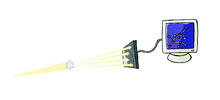
X-ray crystallography
2511
X-ray crystallography allows researchers to see structures too small to be seen by even the most powerful microscopes. Crabtree + Company View Media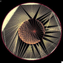
Modeling disease spread
2322
What looks like a Native American dream catcher is really a network of social interactions within a community. Stephen Eubank, University of Virginia Biocomplexity Institute (formerly Virginia Bioinformatics Institute) View Media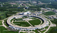
Advanced Photon Source (APS) at Argonne National Lab
2358
The intense X-rays produced by synchrotrons such as the Advanced Photon Source are ideally suited for protein structure determination. Southeast Collaboratory for Structural Genomics View Media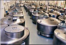
Cryogenic storage tanks at the Coriell Institute for Medical Research
2722
Established in 1953, the Coriell Institute for Medical Research distributes cell lines and DNA samples to researchers around the world. Courtney Sill, Coriell Institute for Medical Research View Media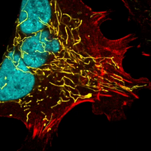
Multinucleated cancer cell
6967
A cancer cell with three nuclei, shown in turquoise. The abnormal number of nuclei indicates that the cell failed to go through cell division, probably more than once. Dylan T. Burnette, Vanderbilt University School of Medicine. View Media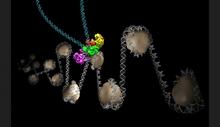
Intasome
6346
Salk researchers captured the structure of a protein complex called an intasome (center) that lets viruses similar to HIV establish permanent infection in their hosts. National Resource for Automated Molecular Microscopy http://nramm.nysbc.org/nramm-images/ Source: Bridget Carragher View MediaAssembly of the HIV capsid
5729
The HIV capsid is a pear-shaped structure that is made of proteins the virus needs to mature and become infective. John Grime and Gregory Voth, The University of Chicago View Media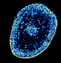
Chromatin in human tenocyte
6893
The nucleus of a degenerating human tendon cell, also known as a tenocyte. It has been color-coded based on the density of chromatin—a substance made up of DNA and proteins. Melike Lakadamyali, Perelman School of Medicine at the University of Pennsylvania. View Media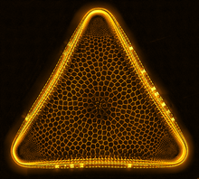
Trigonium diatom
6962
A Trigonium diatom imaged by a quantitative orientation-independent differential interference contrast (OI-DIC) microscope. Michael Shribak, Marine Biological Laboratory/University of Chicago. View Media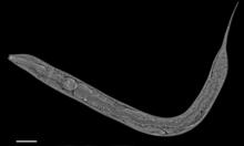
C. elegans showing internal structures
6961
An image of Caenorhabditis elegans, a tiny roundworm, showing internal structures including the intestine, pharynx, and body wall muscle. C. Michael Shribak, Marine Biological Laboratory/University of Chicago. View Media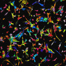
Single-cell “radios” image
7021
Individual cells are color-coded based on their identity and signaling activity using a protein circuit technology developed by the Coyle Lab. Scott Coyle, University of Wisconsin-Madison. View Media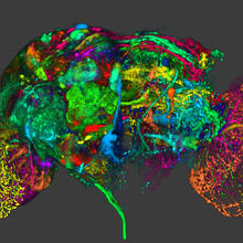
Color coding of the Drosophila brain - image
5838
This image results from a research project to visualize which regions of the adult fruit fly (Drosophila) brain derive from each neural stem cell. Yong Wan from Charles Hansen’s lab, University of Utah. Data preparation and visualization by Masayoshi Ito in the lab of Kei Ito, University of Tokyo. View MediaTracking cells in a gastrulating zebrafish embryo
6776
During development, a zebrafish embryo is transformed from a ball of cells into a recognizable body plan by sweeping convergence and extension cell movements. This process is called gastrulation. Liliana Solnica-Krezel, Washington University School of Medicine in St. Louis. View Media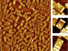
Golden gene chips
2455
A team of chemists and physicists used nanotechnology and DNA's ability to self-assemble with matching RNA to create a new kind of chip for measuring gene activity. Hao Yan and Yonggang Ke, Arizona State University View Media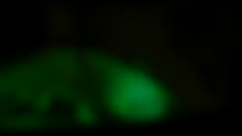
Glow-in-the-dark salamanders
2715
These six-month-old axolotls, a kind of salamander, glow green and blue under ultraviolet light. That's because they were genetically modified to make harmless green fluorescent protein, or GFP. View Media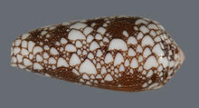
Cone snail shell
2576
A shell from the venomous cone snail Conus omaria, which lives in the Pacific and Indian oceans and eats other snails. Kerry Matz, University of Utah View Media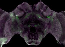
Honeybee brain
6755
Insect brains, like the honeybee brain shown here, are very different in shape from human brains. Gene Robinson, University of Illinois at Urbana-Champaign. View Media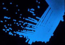
Colorful communication
2313
The marine bacterium Vibrio harveyi glows when near its kind. Bonnie Bassler, Princeton University View Media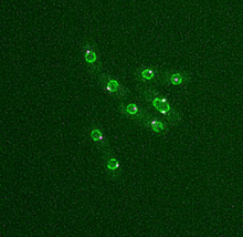
Dividing yeast cells with nuclear envelopes and spindle pole bodies
6795
Time-lapse video of yeast cells undergoing cell division. Nuclear envelopes are shown in green, and spindle pole bodies, which help pull apart copied genetic information, are shown in magenta. Alaina Willet, Kathy Gould’s lab, Vanderbilt University. View Media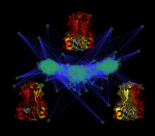
Cluster analysis of mysterious protein
3295
Researchers use cluster analysis to study protein shape and function. Each green circle represents one potential shape of the protein mitoNEET. Patricia Jennings and Elizabeth Baxter, University of California, San Diego View Media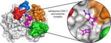
Space-filling model of a cefotaxime-CCD-1 complex
6767
CCD-1 is an enzyme produced by the bacterium Clostridioides difficile that helps it resist antibiotics. Keith Hodgson, Stanford University. View Media
Thermotoga maritima and its metabolic network
2702
A combination of protein structures determined experimentally and computationally shows us the complete metabolic network of a heat-loving bacterium. View Media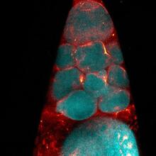
Fruit fly nurse cells during egg development
6753
In many animals, the egg cell develops alongside sister cells. Adam C. Martin, Massachusetts Institute of Technology. View Media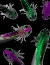
Axolotls showing nervous system components
6928
Axolotls—a type of salamander—that have been genetically modified so that various parts of their nervous systems glow purple and green. Prayag Murawala, MDI Biological Laboratory and Hannover Medical School. View Media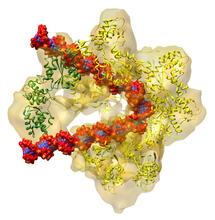
DNA replication origin recognition complex (ORC)
3597
A study published in March 2012 used cryo-electron microscopy to determine the structure of the DNA replication origin recognition complex (ORC), a semi-circular, protein complex (yellow) that recogni Huilin Li, Brookhaven National Laboratory View Media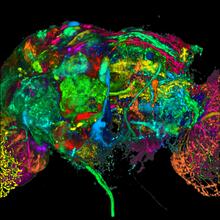
Color coding of the Drosophila brain - black background
5868
This image results from a research project to visualize which regions of the adult fruit fly (Drosophila) brain derive from each neural stem cell. Yong Wan from Charles Hansen’s lab, University of Utah. Data preparation and visualization by Masayoshi Ito in the lab of Kei Ito, University of Tokyo. View Media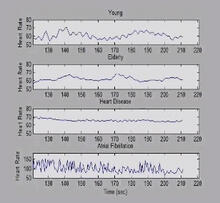
Heart rates time series image
3596
These time series show the heart rates of four different individuals. Madalena Costa and Ary Goldberger, Beth Israel Deaconess Medical Center View Media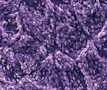
Retinal pigment epithelium derived from human ES cells
3286
This color-enhanced image is a scanning electron microscope image of retinal pigment epithelial (RPE) cells derived from human embryonic stem cells. David Hinton lab, University of Southern California, via CIRM View Media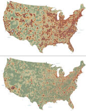
Mapping disease spread
2320
How far and fast an infectious disease spreads across a community depends on many factors, including transportation. These U.S. David Chrest, RTI International View Media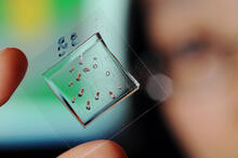
Automated Worm Sorter - 4
3475
Georgia Tech associate professor Hang Lu holds a microfluidic chip that is part of a system that uses artificial intelligence and cutting-edge image processing to automatically examine large number of Georgia Tech/Gary Meek View Media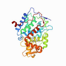
CCP enzyme
6762
The enzyme CCP is found in the mitochondria of baker’s yeast. Scientists study the chemical reactions that CCP triggers, which involve a water molecule, iron, and oxygen. Protein Data Bank. View Media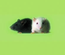
Lab mice
1069
Many researchers use the mouse (Mus musculus) as a model organism to study mammalian biology. Bill Branson, National Institutes of Health View Media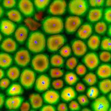
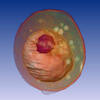
Cell-like compartments from frog eggs
6584
Cell-like compartments that spontaneously emerged from scrambled frog eggs, with nuclei (blue) from frog sperm. Endoplasmic reticulum (red) and microtubules (green) are also visible. Xianrui Cheng, Stanford University School of Medicine. View Media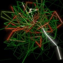
Mapping metabolic activity
2319
Like a map showing heavily traveled roads, this mathematical model of metabolic activity inside an E. coli cell shows the busiest pathway in white. Albert-László Barabási, University of Notre Dame View Media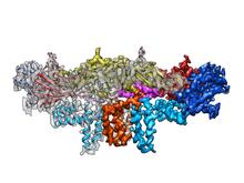
Dengue virus membrane protein structure
3758
Dengue virus is a mosquito-borne illness that infects millions of people in the tropics and subtropics each year. Like many viruses, dengue is enclosed by a protective membrane. Hong Zhou, UCLA View Media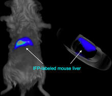
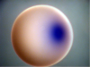
Mouse liver labeled with fluorescent probe
2601
A mouse liver glows after being tagged with specially designed infrared-fluorescent protein (IFP). Xiaokun Shu, University of California, San Diego View Media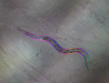
C. elegans trapped by carnivorous fungus
6963
Real-time footage of Caenorhabditis elegans, a tiny roundworm, trapped by a carnivorous fungus, Arthrobotrys dactyloides. Michael Shribak, Marine Biological Laboratory/University of Chicago. View MediaGenetic mosaicism in fruit flies
6983
Fat tissue from the abdomen of a genetically mosaic adult fruit fly. Genetic mosaicism means that the fly has cells with different genotypes even though it formed from a single zygote. Akhila Rajan, Fred Hutchinson Cancer Center View Media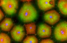
Cell-like compartments from frog eggs 5
6592
Cell-like compartments that spontaneously emerged from scrambled frog eggs, with nuclei (blue) from frog sperm. Endoplasmic reticulum (red) and microtubules (green) are also visible. Xianrui Cheng, Stanford University School of Medicine. View Media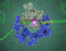
TFIID complex binds DNA to start gene transcription
3766
Gene transcription is a process by which the genetic information encoded in DNA is transcribed into RNA. Eva Nogales, Berkeley Lab View Media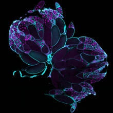
Fruit fly ovaries
6807
Fruit fly (Drosophila melanogaster) ovaries with DNA shown in magenta and actin filaments shown in light blue. This image was captured using a confocal laser scanning microscope.Vladimir I. Gelfand, Feinberg School of Medicine, Northwestern University. View Media
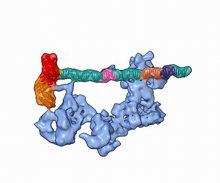
Dynamic cryo-EM model of the human transcription preinitiation complex
5730
Gene transcription is a process by which information encoded in DNA is transcribed into RNA. Eva Nogales, Berkeley Lab View Media