Switch to Gallery View
Image and Video Gallery
This is a searchable collection of scientific photos, illustrations, and videos. The images and videos in this gallery are licensed under Creative Commons Attribution Non-Commercial ShareAlike 3.0. This license lets you remix, tweak, and build upon this work non-commercially, as long as you credit and license your new creations under identical terms.
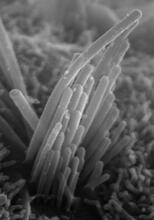
Ear hair cells derived from embryonic stem cells
3272
Mouse embryonic stem cells matured into this bundle of hair cells similar to the ones that transmit sound in the ear. Stefen Heller, Stanford University, via CIRM View Media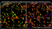
Single-cell “radios” video
7022
Individual cells are color-coded based on their identity and signaling activity using a protein circuit technology developed by the Coyle Lab. Scott Coyle, University of Wisconsin-Madison. View Media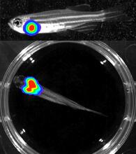
Bioluminescent imaging in adult zebrafish - lateral and overhead view
3556
Luciferase-based imaging enables visualization and quantification of internal organs and transplanted cells in live adult zebrafish. Kenneth Poss, Duke University View Media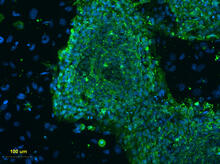
Human embryonic stem cells on feeder cells
3274
This fluorescent microscope image shows human embryonic stem cells whose nuclei are stained green. Blue staining shows the surrounding supportive feeder cells. Michael Longaker lab, Stanford University School of Medicine, via CIRM View Media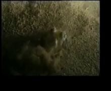
Adult Hawaiian bobtail squid burying in the sand
7012
Each morning, the nocturnal Hawaiian bobtail squid, Euprymna scolopes, hides from predators by digging into the sand. At dusk, it leaves the sand again to hunt. Margaret J. McFall-Ngai, Carnegie Institution for Science/California Institute of Technology, and Edward G. Ruby, California Institute of Technology. View Media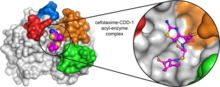
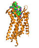
Space-filling model of a cefotaxime-CCD-1 complex
6767
CCD-1 is an enzyme produced by the bacterium Clostridioides difficile that helps it resist antibiotics. Keith Hodgson, Stanford University. View Media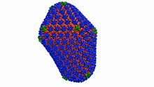
Atomic-level structure of the HIV capsid
6601
This animation shows atoms of the HIV capsid, the shell that encloses the virus's genetic material. Juan R. Perilla and the Theoretical and Computational Biophysics Group, University of Illinois at Urbana-Champaign View Media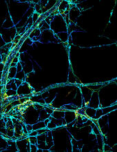
STORM image of axonal cytoskeleton
3678
This image shows the long, branched structures (axons) of nerve cells. Xiaowei Zhuang Laboratory, Howard Hughes Medical Institute, Harvard University View Media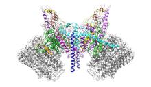
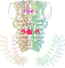
ATP Synthase
6353
Atomic model of the membrane region of the mitochondrial ATP synthase built into a cryo-EM map at 3.6 Å resolution. ATP synthase is the primary producer of ATP in aerobic cells. Bridget Carragher, <a href="http://nramm.nysbc.org/">NRAMM National Resource for Automated Molecular Microscopy</a> View Media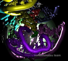
Natural nanomachine in action
2336
Using a supercomputer to simulate the movement of atoms in a ribosome, researchers looked into the core of this protein-making nanomachine and took snapshots. Kevin Sanbonmatsu, Los Alamos National Laboratory View Media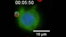
“Two-faced” Janus particle activating a macrophage
6801
A macrophage—a type of immune cell that engulfs invaders—“eats” and is activated by a “two-faced” Janus particle. Yan Yu, Indiana University, Bloomington. View Media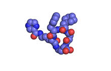
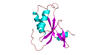
X-ray co-crystal structure of Src kinase bound to a DNA-templated macrocycle inhibitor 1
3413
X-ray co-crystal structure of Src kinase bound to a DNA-templated macrocycle inhibitor. Markus A. Seeliger, Stony Brook University Medical School and David R. Liu, Harvard University View Media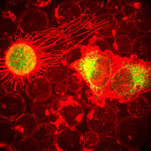
Plasma-Derived Membrane Vesicles
5887
This fiery image doesn’t come from inside a bubbling volcano. Instead, it shows animal cells caught in the act of making bubbles, or blebbing. Jeanne Stachowiak, University of Texas at Austin View Media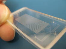
Microfluidic chip
3265
Microfluidic chips have many uses in biology labs. Jeff Hasty Lab, UC San Diego View Media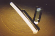
Bovine milk alpha-lactalbumin (2)
2404
Crystals of bovine milk alpha-lactalbumin protein created for X-ray crystallography, which can reveal detailed, three-dimensional protein structures. Alex McPherson, University of California, Irvine View Media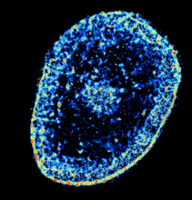
Chromatin in human tenocyte
6893
The nucleus of a degenerating human tendon cell, also known as a tenocyte. It has been color-coded based on the density of chromatin—a substance made up of DNA and proteins. Melike Lakadamyali, Perelman School of Medicine at the University of Pennsylvania. View Media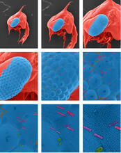
Crab larva eye
1251
Colorized scanning electron micrographs progressively zoom in on the eye of a crab larva. In the higher-resolution frames, bacteria are visible on the eye. Tina Weatherby Carvalho, University of Hawaii at Manoa View Media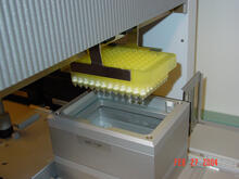
Protein purification robot in action 01
2369
A robot is transferring 96 purification columns to a vacuum manifold for subsequent purification procedures. The Northeast Collaboratory for Structural Genomics View Media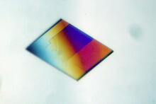
Bacterial alpha amylase
2401
A crystal of bacterial alpha amylase protein created for X-ray crystallography, which can reveal detailed, three-dimensional protein structures. Alex McPherson, University of California, Irvine View Media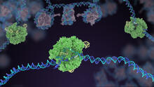
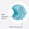
Cas9 protein involved in the CRISPR gene-editing technology
5816
In the gene-editing tool CRISPR, a small strand of RNA identifies a specific chunk of DNA. Janet Iwasa View Media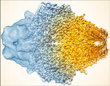
Beta-galactosidase montage showing cryo-EM improvement--gradient background
5883
Composite image of beta-galactosidase showing how cryo-EM’s resolution has improved dramatically in recent years. Older images to the left, more recent to the right. Veronica Falconieri, Sriram Subramaniam Lab, National Cancer Institute View Media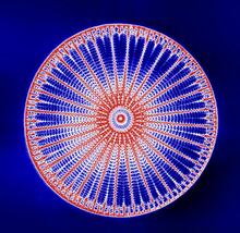
Arachnoidiscus diatom
6902
An Arachnoidiscus diatom with a diameter of 190µm. Michael Shribak, Marine Biological Laboratory/University of Chicago. View Media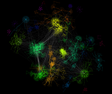
A molecular interaction network in yeast 2
3732
The image visualizes a part of the yeast molecular interaction network. Keiichiro Ono, UCSD View Media
Scientists display X-ray diffraction pattern obtained with split X-ray beamline
2384
Scientists from Argonne National Laboratory's Advanced Photon Source (APS) display the first X-ray diffraction pattern obtained from a protein crystal using a split X-ray beam, the first of its kind a GM/CA Collaborative Access Team View Media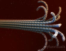
Microtubule breakdown
2321
Like a building supported by a steel frame, a cell contains its own sturdy internal scaffolding made up of proteins, including microtubules. Eva Nogales, University of California, Berkeley View Media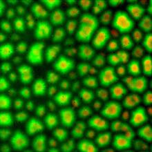
Cell-like compartments from frog eggs 3
6586
Cell-like compartments that spontaneously emerged from scrambled frog eggs. Endoplasmic reticulum (red) and microtubules (green) are visible. Image created using epifluorescence microscopy. Xianrui Cheng, Stanford University School of Medicine. View Media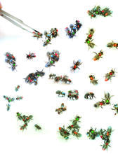
Honeybees marked with paint
6756
Researchers doing behavioral experiments with honeybees sometimes use paint or enamel to give individual bees distinguishing marks. Gene Robinson, University of Illinois at Urbana-Champaign. View Media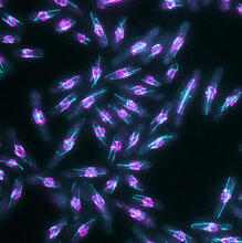
Yeast cells with nuclear envelopes and tubulin
6798
Yeast cells with nuclear envelopes shown in magenta and tubulin shown in light blue. The nuclear envelope defines the borders of the nucleus, which houses DNA. Alaina Willet, Kathy Gould’s lab, Vanderbilt University. View Media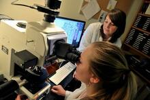
Research mentor and student
2767
A research mentor (Lori Eidson) and student (Nina Waldron, on the microscope) were 2009 members of the BRAIN (Behavioral Research Advancements In Neuroscience) program at Georgia State University in A Elizabeth Weaver, Georgia State University View Media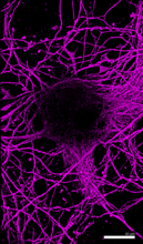
Microtubules in hippocampal neurons
6890
Microtubules (magenta) in neurons of the hippocampus, a part of the brain involved in learning and memory. Microtubules are strong, hollow fibers that provide structural support to cells. Melike Lakadamyali, Perelman School of Medicine at the University of Pennsylvania. View Media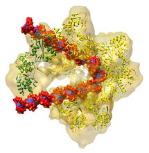
DNA replication origin recognition complex (ORC)
3597
A study published in March 2012 used cryo-electron microscopy to determine the structure of the DNA replication origin recognition complex (ORC), a semi-circular, protein complex (yellow) that recogni Huilin Li, Brookhaven National Laboratory View Media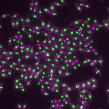
Yeast cells with nuclei and contractile rings
6792
Yeast cells with nuclei shown in green and contractile rings shown in magenta. Nuclei store DNA, and contractile rings help cells divide. Alaina Willet, Kathy Gould’s lab, Vanderbilt University. View Media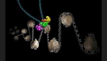
Intasome
6346
Salk researchers captured the structure of a protein complex called an intasome (center) that lets viruses similar to HIV establish permanent infection in their hosts. National Resource for Automated Molecular Microscopy http://nramm.nysbc.org/nramm-images/ Source: Bridget Carragher View Media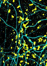
Lysosomes and microtubules
6889
Lysosomes (yellow) and detyrosinated microtubules (light blue). Lysosomes are bubblelike organelles that take in molecules and use enzymes to break them down. Melike Lakadamyali, Perelman School of Medicine at the University of Pennsylvania. View Media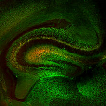
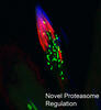
Mouse Brain Cross Section
5886
The brain sections are treated with fluorescent antibodies specific to a particular protein and visualized using serial electron microscopy (SEM). Anton Maximov, The Scripps Research Institute, La Jolla, CA View Media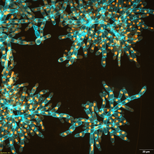
Snowflake yeast 3
6971
Multicellular yeast called snowflake yeast that researchers created through many generations of directed evolution from unicellular yeast. William Ratcliff, Georgia Institute of Technology. View Media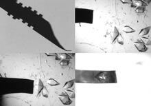
Mounting of protein crystals
2368
Automated methods using micromachined silicon are used at the Northeast Collaboratory for Structural Genomics to mount protein crystals for X-ray crystallography. The Northeast Collaboratory for Structural Genomics View Media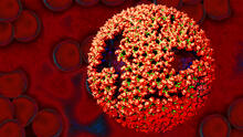
Molecular model of freshly made Rous sarcoma virus (RSV)
3771
Viruses have been the foes of animals and other organisms for time immemorial. Boon Chong Goh, University of Illinois at Urbana-Champaign View Media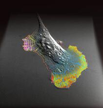
Biosensors illustration
2802
A rendering of an activity biosensor image overlaid with a cell-centered frame of reference used for image analysis of signal transduction. Gaudenz Danuser, Harvard Medical School View Media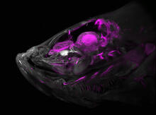
Zebrafish head vasculature
6934
A zebrafish head with blood vessels shown in purple. Prayag Murawala, MDI Biological Laboratory and Hannover Medical School. View Media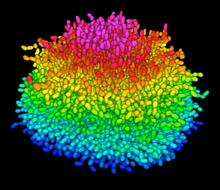
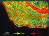
A Growing Bacterial Biofilm
5825
A growing Vibrio cholerae (cholera) biofilm. Cholera bacteria form colonies called biofilms that enable them to resist antibiotic therapy within the body and other challenges to their growth. Jing Yan, Ph.D., and Bonnie Bassler, Ph.D., Department of Molecular Biology, Princeton University, Princeton, NJ. View Media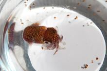
Adult and juvenile Hawaiian bobtail squids
7010
An adult Hawaiian bobtail squid, Euprymna scolopes, (~4 cm) surrounded by newly hatched juveniles (~2 mm) in a bowl of seawater.Margaret J. McFall-Ngai, Carnegie Institution for Science/California Institute of Technology, and Edward G. Ruby, California Institute of Technology. View Media
Fruit fly spermatids
3590
Developing spermatids (precursors of mature sperm cells) begin as small, round cells and mature into long-tailed, tadpole-shaped ones. Lacramioara Fabian, The Hospital for Sick Children, Toronto, Canada View Media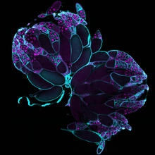
Fruit fly ovaries
6807
Fruit fly (Drosophila melanogaster) ovaries with DNA shown in magenta and actin filaments shown in light blue. This image was captured using a confocal laser scanning microscope.Vladimir I. Gelfand, Feinberg School of Medicine, Northwestern University. View Media
Cryo-electron microscopy revealing the "wasabi receptor"
3747
The TRPA1 protein is responsible for the burn you feel when you taste a bite of sushi topped with wasabi. Jean-Paul Armache, UCSF View Media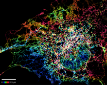
Dense tubular matrices in the peripheral endoplasmic reticulum (ER) 1
5855
Superresolution microscopy work on endoplasmic reticulum (ER) in the peripheral areas of the cell showing details of the structure and arrangement in a complex web of tubes. Jennifer Lippincott-Schwartz, Howard Hughes Medical Institute Janelia Research Campus, Virginia View Media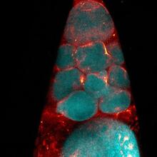
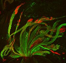
Fruit fly nurse cells during egg development
6753
In many animals, the egg cell develops alongside sister cells. Adam C. Martin, Massachusetts Institute of Technology. View Media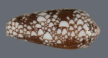
Cone snail shell
2576
A shell from the venomous cone snail Conus omaria, which lives in the Pacific and Indian oceans and eats other snails. Kerry Matz, University of Utah View Media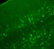
Confocal microscopy of perineuronal nets in the brain 2
3742
The photo shows a confocal microscopy image of perineuronal nets (PNNs), which are specialized extracellular matrix (ECM) structures in the brain. Tom Deerinck, National Center for Microscopy and Imaging Research (NCMIR) View Media
Vimentin in a quail embryo
2809
Video of high-resolution confocal images depicting vimentin immunofluorescence (green) and nuclei (blue) at the edge of a quail embryo yolk. Andrés Garcia, Georgia Tech View Media