Switch to Gallery View
Image and Video Gallery
This is a searchable collection of scientific photos, illustrations, and videos. The images and videos in this gallery are licensed under Creative Commons Attribution Non-Commercial ShareAlike 3.0. This license lets you remix, tweak, and build upon this work non-commercially, as long as you credit and license your new creations under identical terms.
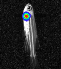
Bioluminescent imaging in adult zebrafish - overhead view
3557
Luciferase-based imaging enables visualization and quantification of internal organs and transplanted cells in live adult zebrafish. Kenneth Poss, Duke University View Media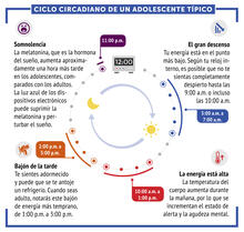
Ciclo circadiano de un adolescente típico
6612
Los ritmos circadianos son cambios físicos, mentales y conductuales que siguen un ciclo de 24 horas. NIGMS View Media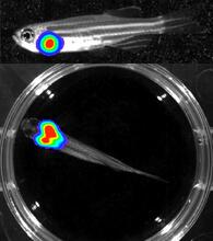
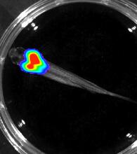
Bioluminescent imaging in adult zebrafish - lateral and overhead view
3556
Luciferase-based imaging enables visualization and quantification of internal organs and transplanted cells in live adult zebrafish. Kenneth Poss, Duke University View Media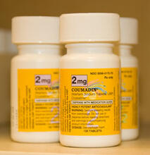
Bottles of warfarin
2579
In 2007, the FDA modified warfarin's label to indicate that genetic makeup may affect patient response to the drug. The widely used blood thinner is sold under the brand name Coumadin®. Alisa Machalek, NIGMS/NIH View Media
Movie of in vitro assembly of a cell-signaling pathway
3786
T cells are white blood cells that are important in defending the body against bacteria, viruses and other pathogens. Xiaolei Su, HHMI Whitman Center of the Marine Biological Laboratory View Media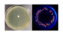
Antibiotic-surviving bacteria
6802
Colonies of bacteria growing despite high concentrations of antibiotics. These colonies are visible both by eye, as seen on the left, and by bioluminescence imaging, as seen on the right. Paul Stoodley, The Ohio State University. View Media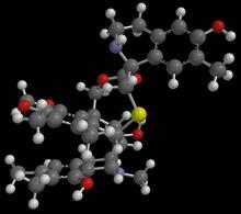
Anti-tumor drug ecteinascidin 743 (ET-743) with hydrogens 01
2790
Ecteinascidin 743 (ET-743, brand name Yondelis), was discovered and isolated from a sea squirt, Ecteinascidia turbinata, by NIGMS grantee Kenneth Rinehart at the University of Illinois. Timothy Jamison, Massachusetts Institute of Technology View Media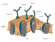
Plasma membrane (with labels)
2524
The plasma membrane is a cell's protective barrier. See image 2523 for an unlabeled version of this illustration. Featured in The Chemistry of Health. Crabtree + Company View Media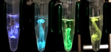
Bioluminescence in a Tube
5895
Details about the basic biology and chemistry of the ingredients that produce bioluminescence are allowing scientists to harness it as an imaging tool. Credit: Nathan Shaner, Scintillon Institute. Nathan Shaner, Scintillon Institute View Media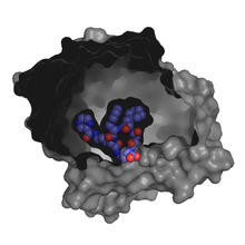
X-ray co-crystal structure of Src kinase bound to a DNA-templated macrocycle inhibitor 3
3415
X-ray co-crystal structure of Src kinase bound to a DNA-templated macrocycle inhibitor. Markus A. Seeliger, Stony Brook University Medical School and David R. Liu, Harvard University View Media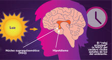
Los ritmos circadianos y el núcleo supraquiasmático
6614
Los ritmos circadianos son cambios físicos, mentales y de comportamiento que siguen un ciclo de 24 horas. NIGMS View Media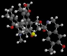
Anti-tumor drug ecteinascidin 743 (ET-743) with hydrogens 03
2792
Ecteinascidin 743 (ET-743, brand name Yondelis), was discovered and isolated from a sea squirt, Ecteinascidia turbinata, by NIGMS grantee Kenneth Rinehart at the University of Illinois. Timothy Jamison, Massachusetts Institute of Technology View Media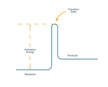
Activation energy (with labels)
2526
To become products, reactants must overcome an energy hill. See image 2525 for an unlabeled version of this illustration. Crabtree + Company View Media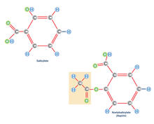
Aspirin (with labels)
2530
Acetylsalicylate (bottom) is the aspirin of today. Crabtree + Company View Media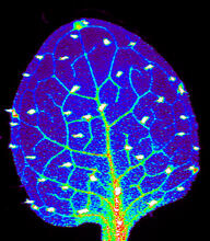
Zinc levels in a plant leaf
3727
Zinc is required for the function of more than 300 enzymes, including those that help regulate gene expression, in various organisms including humans. Suzana Car, Dartmouth College View Media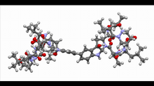
Himastatin, 360-degree view
6851
A 360-degree view of the molecule himastatin, which was first isolated from the bacterium Streptomyces himastatinicus. Himastatin shows antibiotic activity. Mohammad Movassaghi, Massachusetts Institute of Technology. View Media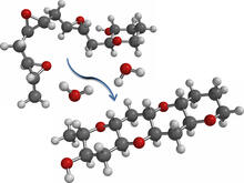
Cascade reaction promoted by water
2490
This illustration of an epoxide-opening cascade promoted by water emulates the proposed biosynthesis of some of the Red Tide toxins. Tim Jamison, Massachusetts Institute of Technology View Media
X-ray co-crystal structure of Src kinase bound to a DNA-templated macrocycle inhibitor 5
3417
X-ray co-crystal structure of Src kinase bound to a DNA-templated macrocycle inhibitor. Markus A. Seeliger, Stony Brook University Medical School and David R. Liu, Harvard University View Media
Anti-tumor drug ecteinascidin 743 (ET-743) with hydrogens 02
2791
Ecteinascidin 743 (ET-743, brand name Yondelis), was discovered and isolated from a sea squirt, Ecteinascidia turbinata, by NIGMS grantee Kenneth Rinehart at the University of Illinois. Timothy Jamison, Massachusetts Institute of Technology View Media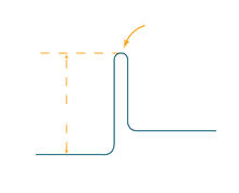
Activation energy
2525
To become products, reactants must overcome an energy hill. See image 2526 for a labeled version of this illustration. Featured in The Chemistry of Health. Crabtree + Company View Media
Antibodies in silica honeycomb
2750
Antibodies are among the most promising therapies for certain forms of cancer, but patients must take them intravenously, exposing healthy tissues to the drug and increasing the risk of side effects. Chenghong Lei, Pacific Northwest National Laboratory & Karl Erik Hellstrom, University of Washington View Media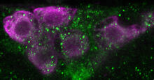
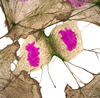
Insulin production and fat sensing in fruit flies
6982
Fourteen neurons (magenta) in the adult Drosophila brain produce insulin, and fat tissue sends packets of lipids to the brain via the lipoprotein carriers (green). Akhila Rajan, Fred Hutchinson Cancer Center View Media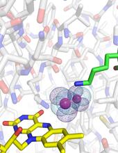
O2 reacting with a flavin-dependent enzyme
3411
Department of Biological Chemistry, University of Michigan View Media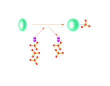
Kinases
2534
Kinases are enzymes that add phosphate groups (red-yellow structures) to proteins (green), assigning the proteins a code. Crabtree + Company View Media
Bacillus anthracis being killed
3525
Bacillus anthracis (anthrax) cells being killed by a fluorescent trans-translation inhibitor, which disrupts bacterial protein synthesis. Kenneth Keiler, Penn State University View Media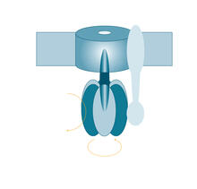
ATP synthase
2517
The world's smallest motor, ATP synthase, generates energy for the cell. See image 2518 for a labeled version of this illustration. Crabtree + Company View Media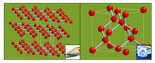
Carbon building blocks (with examples)
2507
The arrangement of identical molecular components can make a dramatic difference. For example, carbon atoms can be arranged into dull graphite (left) or sparkly diamonds (right). Crabtree + Company View Media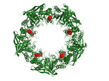
Cas4 nuclease protein structure
3720
This wreath represents the molecular structure of a protein, Cas4, which is part of a system, known as CRISPR, that bacteria use to protect themselves against viral invaders. Fred Dyda, NIDDK View Media
Genetically identical mycobacteria respond differently to antibiotic 2
5752
Antibiotic resistance in microbes is a serious health concern. So researchers have turned their attention to how bacteria undo the action of some antibiotics. Bree Aldridge, Tufts University View Media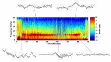
Brain waves of a patient anesthetized with propofol
6779
A representation of a patient’s brain waves after receiving the anesthetic propofol. Emery N. Brown, M.D., Ph.D., Massachusetts General Hospital/Harvard Medical School, Picower Institute for Learning and Memory, and Massachusetts Institute of Technology. View Media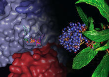
Chang Shan
3483
For thousands of years, Chinese herbalists have treated malaria using Chang Shan, a root extract from a type of hydrangea that grows in Tibet and Nepal. Paul Schimmel Lab, Scripps Research Institute View Media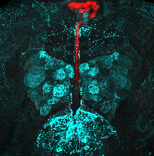
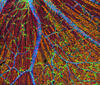
Fruit fly brain responds to adipokines
6985
Drosophila adult brain showing that an adipokine (fat hormone) generates a response from neurons (aqua) and regulates insulin-producing neurons (red).Akhila Rajan, Fred Hutchinson Cancer Center View Media
Bioluminescent imaging in adult zebrafish - lateral view
3558
Luciferase-based imaging enables visualization and quantification of internal organs and transplanted cells in live adult zebrafish. Kenneth Poss, Duke University View Media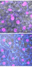
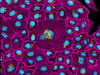
Fruit fly starvation leads to adipokine accumulation
6984
Adult Drosophila abdominal fat tissue showing cell nuclei labelled in magenta. Akhila Rajan, Fred Hutchinson Cancer Center View Media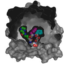
X-ray co-crystal structure of Src kinase bound to a DNA-templated macrocycle inhibitor 4
3416
X-ray co-crystal structure of Src kinase bound to a DNA-templated macrocycle inhibitor. Markus A. Seeliger, Stony Brook University Medical School and David R. Liu, Harvard University View Media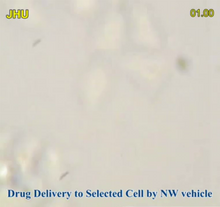
Precisely Delivering Chemical Cargo to Cells
3779
Moving protein or other molecules to specific cells to treat or examine them has been a major biological challenge. Nature Nanotechnology View Media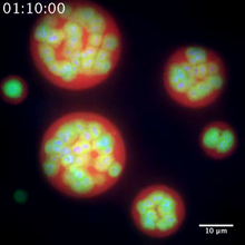
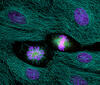
Nucleolus subcompartments spontaneously self-assemble 2
3791
The nucleolus is a small but very important protein complex located in the cell's nucleus. Nilesh Vaidya, Princeton University View Media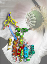
Atomic Structure of Poppy Enzyme
3422
The atomic structure of the morphine biosynthetic enzyme salutaridine reductase bound to the cofactor NADPH. The substrate salutaridine is shown entering the active site. Judy Coyle, Donald Danforth Plant Science Center View Media
Nucleolus subcompartments spontaneously self-assemble 1
3789
The nucleolus is a small but very important protein complex located in the cell's nucleus. Nilesh Vaidya, Princeton University View Media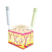
Drugs enter skin
2531
Drugs enter different layers of skin via intramuscular, subcutaneous, or transdermal delivery methods. See image 2532 for a labeled version of this illustration. Crabtree + Company View Media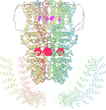
Cryo-electron microscopy revealing the "wasabi receptor"
3747
The TRPA1 protein is responsible for the burn you feel when you taste a bite of sushi topped with wasabi. Jean-Paul Armache, UCSF View Media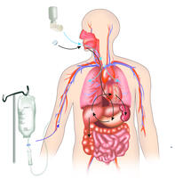
A drug's life in the body
2527
A drug's life in the body. Medicines taken by mouth pass through the liver before they are absorbed into the bloodstream. Crabtree + Company View Media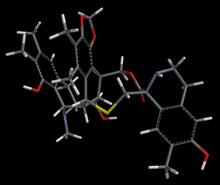
Anti-tumor drug ecteinascidin 743 (ET-743) with hydrogens 04
2793
Ecteinascidin 743 (ET-743, brand name Yondelis), was discovered and isolated from a sea squirt, Ecteinascidia turbinata, by NIGMS grantee Kenneth Rinehart at the University of Illinois. Timothy Jamison, Massachusetts Institute of Technology View Media
HIV Capsid
3477
This image is a computer-generated model of the approximately 4.2 million atoms of the HIV capsid, the shell that contains the virus' genetic material. Juan R. Perilla and the Theoretical and Computational Biophysics Group, University of Illinois at Urbana-Champaign View Media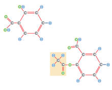

A drug's life in the body (with labels)
2528
A drug's life in the body. Medicines taken by mouth (oral) pass through the liver before they are absorbed into the bloodstream. Crabtree + Company View Media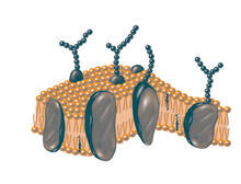
Plasma membrane
2523
The plasma membrane is a cell's protective barrier. See image 2524 for a labeled version of this illustration. Featured in The Chemistry of Health. Crabtree + Company View Media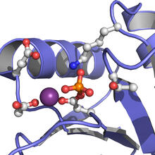
Active Site of E. coli response regulator PhoB
3412
Active site of E. coli response regulator PhoB. Ann Stock, Rutgers University View Media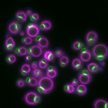
Yeast cells responding to a glucose shortage
6772
These yeast cells were exposed to a glucose (sugar) shortage. Mike Henne, University of Texas Southwestern Medical Center. View Media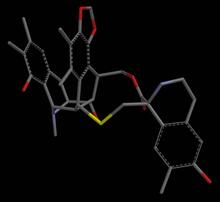
Anti-tumor drug ecteinascidin 743 (ET-743), structure without hydrogens 04
2797
Ecteinascidin 743 (ET-743, brand name Yondelis), was discovered and isolated from a sea squirt, Ecteinascidia turbinata, by NIGMS grantee Kenneth Rinehart at the University of Illinois. Timothy Jamison, Massachusetts Institute of Technology View Media