Switch to Gallery View
Image and Video Gallery
This is a searchable collection of scientific photos, illustrations, and videos. The images and videos in this gallery are licensed under Creative Commons Attribution Non-Commercial ShareAlike 3.0. This license lets you remix, tweak, and build upon this work non-commercially, as long as you credit and license your new creations under identical terms.
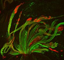
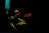
Fruit fly spermatids
3590
Developing spermatids (precursors of mature sperm cells) begin as small, round cells and mature into long-tailed, tadpole-shaped ones. Lacramioara Fabian, The Hospital for Sick Children, Toronto, Canada View Media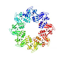
Protein involved in cell division from Mycoplasma pneumoniae
2377
Model of a protein involved in cell division from Mycoplasma pneumoniae. This model, based on X-ray crystallography, revealed a structural domain not seen before. Berkeley Structural Genomics Center, PSI View Media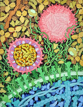
Zika virus
6998
Zika virus is shown in cross section at center left. On the outside, it includes envelope protein (red) and membrane protein (magenta) embedded in a lipid membrane (light purple). Amy Wu and Christine Zardecki, RCSB Protein Data Bank. View Media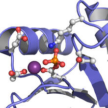
Active Site of E. coli response regulator PhoB
3412
Active site of E. coli response regulator PhoB. Ann Stock, Rutgers University View Media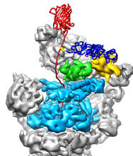
The 26S proteasome engages with a protein substrate
3763
The proteasome is a critical multiprotein complex in the cell that breaks down and recycles proteins that have become damaged or are no longer needed. Andreas Martin, HHMI View Media
Human endoplasmic reticulum membrane protein complex
6777
A 3D model of the human endoplasmic reticulum membrane protein complex (EMC) that identifies its nine essential subunits. Rebecca Voorhees, California Institute of Technology. View Media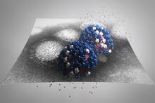
Stetten Lecture 2017poster image
5896
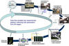
This image is featured on the poster for Dr. Rommie Amaro's 2017 Stetten Lecture. Dr. Rommie Amaro, University of California, San Diego View MediaNuclear Lamina – Three Views
6573
Three views of the entire nuclear lamina of a HeLa cell produced by tilted light sheet 3D single-molecule super-resolution imaging using a platform termed TILT3D. Anna-Karin Gustavsson, Ph.D. View Media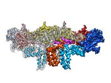
Dengue virus membrane protein structure
3758
Dengue virus is a mosquito-borne illness that infects millions of people in the tropics and subtropics each year. Like many viruses, dengue is enclosed by a protective membrane. Hong Zhou, UCLA View Media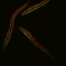
Group of fluorescent C. elegans showing muscle and ribosomal protein
6582
Three C. elegans, tiny roundworms, with a ribosomal protein glowing red and muscle fibers glowing green. Researchers used these worms to study a molecular pathway that affects aging. Jarod Rollins, Mount Desert Island Biological Laboratory. View Media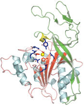
Human aspartoacylase
2352
Model of aspartoacylase, a human enzyme involved in brain metabolism. Center for Eukaryotic Structural Genomics, PSI View Media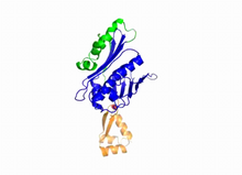
Hsp33 Heat Shock Protein Inactive to Active
3402
When the heat shock protein hsp33 is folded, it is inactive and contains a zinc ion, stabilizing the redox sensitive domain (orange). Dana Reichmann, University of Michigan View Media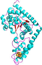
Trp_RS - tryptophanyl tRNA-synthetase family of enzymes
2483
This image represents the structure of TrpRS, a novel member of the tryptophanyl tRNA-synthetase family of enzymes. View Media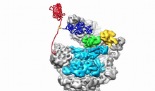
Movie of the 19S proteasome subunit processing a protein substrate
3764
The proteasome is a critical multiprotein complex in the cell that breaks down and recycles proteins that have become damaged or are no longer needed. Andreas Martin, HHMI View Media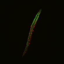
Fluorescent C. elegans showing muscle and ribosomal protein
6581
C. elegans, a tiny roundworm, with a ribosomal protein glowing red and muscle fibers glowing green. Researchers used these worms to study a molecular pathway that affects aging. Jarod Rollins, Mount Desert Island Biological Laboratory. View Media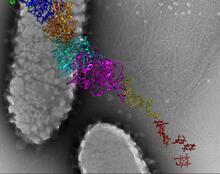
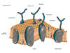
Bacterial nanowire model
6580
A model of a Geobacter sulfurreducens nanowire created from cryo-electron microscopy images. Edward Egelman, University of Virginia. View Media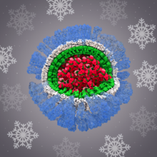
H1N1 Influenza Virus
6356
Related to image 6355. Dr. Rommie Amaro, University of California, San Diego View Media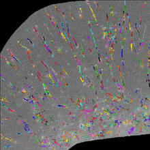
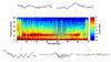
Trajectories of labeled cell receptors
2801
Trajectories of single molecule labeled cell surface receptors. This is an example of NIH-supported research on single-cell analysis. Gaudenz Danuser, Harvard Medical School View Media