Switch to Gallery View
Image and Video Gallery
This is a searchable collection of scientific photos, illustrations, and videos. The images and videos in this gallery are licensed under Creative Commons Attribution Non-Commercial ShareAlike 3.0. This license lets you remix, tweak, and build upon this work non-commercially, as long as you credit and license your new creations under identical terms.
Wound healing in process
3500
Wound healing requires the action of stem cells. Hermann Steller, Rockefeller University View Media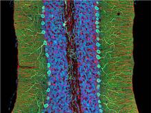
Mouse cerebellum close-up
3371
The cerebellum is the brain's locomotion control center. Every time you shoot a basketball, tie your shoe or chop an onion, your cerebellum fires into action. National Center for Microscopy and Imaging Research (NCMIR) View Media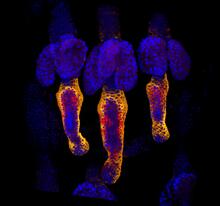
Growing hair follicle stem cells
3499
Wound healing requires the action of stem cells. Hermann Steller, Rockefeller University View Media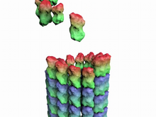
How a microtubule builds and deconstructs
3650
A microtubule, part of the cell's skeleton, builds and deconstructs. View Media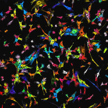
Single-cell “radios” image
7021
Individual cells are color-coded based on their identity and signaling activity using a protein circuit technology developed by the Coyle Lab. Scott Coyle, University of Wisconsin-Madison. View Media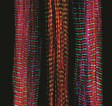
Three muscle fibers; the middle has a defect found in some neuromuscular diseases
3630
Of the three muscle fibers shown here, the one on the right and the one on the left are normal. The middle fiber is deficient a large protein called nebulin (blue). Christopher Pappas and Carol Gregorio, University of Arizona View Media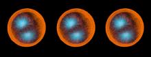
Sea urchin embryo 02
1048
Stereo triplet of a sea urchin embryo stained to reveal actin filaments (orange) and microtubules (blue). George von Dassow, University of Washington View Media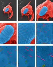
Crab larva eye
1251
Colorized scanning electron micrographs progressively zoom in on the eye of a crab larva. In the higher-resolution frames, bacteria are visible on the eye. Tina Weatherby Carvalho, University of Hawaii at Manoa View Media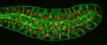
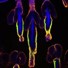
Salivary gland in the developing fruit fly
3603
For fruit flies, the salivary gland is used to secrete materials for making the pupal case, the protective enclosure in which a larva transforms into an adult fly. Richard Fehon, University of Chicago View Media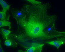
Smooth muscle from human ES cells
3288
These smooth muscle cells were derived from human embryonic stem cells. The nuclei are stained blue, and the proteins of the cytoskeleton are stained green. Alexey Terskikh lab, Burnham Institute for Medical Research, via CIRM View Media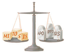
Life in balance
1336
Mitosis creates cells, and apoptosis kills them. The processes often work together to keep us healthy. Judith Stoffer View Media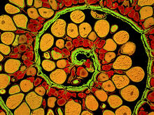
Anglerfish ovary cross-section
3620
This image captures the spiral-shaped ovary of an anglerfish in cross-section. Once matured, these eggs will be released in a gelatinous, floating mass. James E. Hayden, The Wistar Institute, Philadelphia, Pa. View Media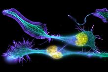
Developing nerve cells
3632
These developing mouse nerve cells have a nucleus (yellow) surrounded by a cell body, with long extensions called axons and thin branching structures called dendrites. Torsten Wittmann, University of California, San Francisco View Media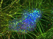
Peripheral nerve cell derived from ES cells
3264
A peripheral nerve cell made from human embryonic stem cell-derived neural crest stem cells. Stephen Dalton, University of Georgia View Media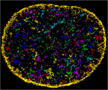
Chromatin in human fibroblast
6888
The nucleus of a human fibroblast cell with chromatin—a substance made up of DNA and proteins—shown in various colors. Melike Lakadamyali, Perelman School of Medicine at the University of Pennsylvania. View Media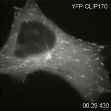
Microtubule dynamics in real time
2784
Cytoplasmic linker protein (CLIP)-170 is a microtubule plus-end-tracking protein that regulates microtubule dynamics and links microtubule ends to different intracellular structures. Gary Borisy, Marine Biology Laboratory View Media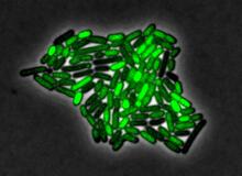
Pulsating response to stress in bacteria
3253
By attaching fluorescent proteins to the genetic circuit responsible for B. subtilis's stress response, researchers can observe the cells' pulses as green flashes. Michael Elowitz, Caltech University View Media
Human endoplasmic reticulum membrane protein complex
6777
A 3D model of the human endoplasmic reticulum membrane protein complex (EMC) that identifies its nine essential subunits. Rebecca Voorhees, California Institute of Technology. View Media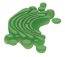
Golgi
1275
The Golgi complex, also called the Golgi apparatus or, simply, the Golgi. Judith Stoffer View Media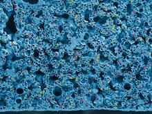
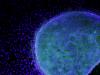
Yeast cells pack a punch
3788
Although they are tiny, microbes that are growing in confined spaces can generate a lot of pressure. In this video, yeast cells grow in a small chamber called a microfluidic bioreactor. Oskar Hallatschek, UC Berkeley View Media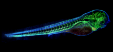
Zebrafish embryo showing vasculature
6661
A zebrafish embryo. The blue areas are cell bodies, the green lines are blood vessels, and the red glow is blood. Kevin Eliceiri, University of Wisconsin-Madison. View Media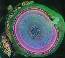
A mammalian eye has approximately 70 different cell types
3641
The incredible complexity of a mammalian eye (in this case from a mouse) is captured here. Each color represents a different type of cell. Bryan William Jones and Robert E. Marc, University of Utah View Media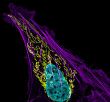
Bone cancer cell
3626
This image shows an osteosarcoma cell with DNA in blue, energy factories (mitochondria) in yellow, and actin filaments—part of the cellular skeleton—in purple. Dylan Burnette and Jennifer Lippincott-Schwartz, NICHD View Media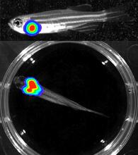
Bioluminescent imaging in adult zebrafish - lateral and overhead view
3556
Luciferase-based imaging enables visualization and quantification of internal organs and transplanted cells in live adult zebrafish. Kenneth Poss, Duke University View Media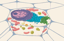
Animal cell
1274
A typical animal cell, sliced open to reveal a cross-section of organelles. Judith Stoffer View Media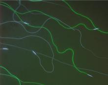
GFP sperm
2683
Fruit fly sperm cells glow bright green when they express the gene for green fluorescent protein (GFP). View Media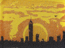
Yeast art depicting the New York City skyline
6521
This skyline of New York City was created by “printing” nanodroplets containing yeast (Saccharomyces cerevisiae) onto a large plate. Each dot is a separate yeast colony. Michael Shen, Ph.D., Jasmine Temple, Leslie Mitchell, Ph.D., and Jef Boeke, Ph.D., New York University School of Medicine; and Nick Phillips, James Chuang, Ph.D., and Jiarui Wang, Johns Hopkins University. View Media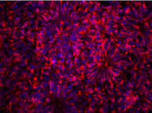
Neurons from human ES cells
3284
These neural precursor cells were derived from human embryonic stem cells. The neural cell bodies are stained red, and the nuclei are blue. Xianmin Zeng lab, Buck Institute for Age Research, via CIRM View Media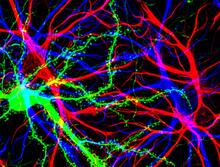
Brain cells in the hippocampus
3688
Hippocampal cells in culture with a neuron in green, showing hundreds of the small protrusions known as dendritic spines. Shelley Halpain, UC San Diego View Media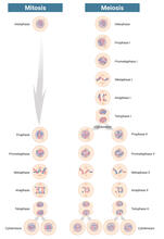
Mitosis and meiosis compared-labeled
6788
Meiosis is used to make sperm and egg cells. During meiosis, a cell's chromosomes are copied once, but the cell divides twice. Judith Stoffer View Media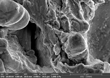
Staphylococcus aureus in the porous coating of a femoral hip stem
6804
Staphylococcus aureus bacteria (blue) on the porous coating of a femoral hip stem used in hip replacement surgery. Paul Stoodley, The Ohio State University. View Media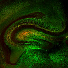
Mouse Brain Cross Section
5886
The brain sections are treated with fluorescent antibodies specific to a particular protein and visualized using serial electron microscopy (SEM). Anton Maximov, The Scripps Research Institute, La Jolla, CA View Media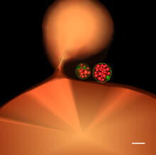
Multivesicular bodies containing intralumenal vesicles assemble at the vacuole 1
5769
Collecting and transporting cellular waste and sorting it into recylable and nonrecylable pieces is a complex business in the cell. Matthew West and Greg Odorizzi, University of Colorado View Media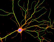
Hippocampal neuron in culture
3687
Hippocampal neuron in culture. Dendrites are green, dendritic spines are red and DNA in cell's nucleus is blue. Shelley Halpain, UC San Diego View Media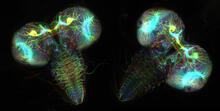
Fruit fly larvae brains showing tubulin
6808
Two fruit fly (Drosophila melanogaster) larvae brains with neurons expressing fluorescently tagged tubulin protein. Vladimir I. Gelfand, Feinberg School of Medicine, Northwestern University. View Media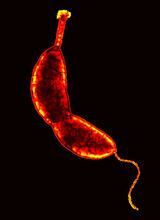
Caulobacter
3262
A study using Caulobacter crescentus showed that some bacteria use just-in-time processing, much like that used in industrial delivery, to make the glue that allows them to attach to surfaces, Yves Brun, Indiana University View Media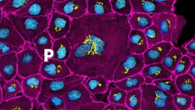
Pathways: The Fascinating Cells of Research Organisms
6538
Learn how research organisms, such as fruit flies and mice, can help us understand and treat human diseases. National Institute of General Medical Sciences View Media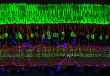
The eye uses many layers of nerve cells to convert light into sight
3635
This image captures the many layers of nerve cells in the retina. The top layer (green) is made up of cells called photoreceptors that convert light into electrical signals to relay to the brain. Wei Li, National Eye Institute, National Institutes of Health View Media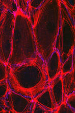
Cells lining the blood vessel walls
3633
The structure of the endothelium, the thin layer of cells that line our arteries and veins, is visible here. Christopher V. Carman and Roberta Martinelli, Harvard Medical School. View Media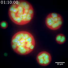
Nucleolus subcompartments spontaneously self-assemble 2
3791
The nucleolus is a small but very important protein complex located in the cell's nucleus. Nilesh Vaidya, Princeton University View Media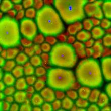
Cell-like compartments from frog eggs 2
6585
Cell-like compartments that spontaneously emerged from scrambled frog eggs, with nuclei (blue) from frog sperm. Endoplasmic reticulum (red) and microtubules (green) are also visible. Xianrui Cheng, Stanford University School of Medicine. View Media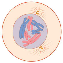
Mitosis - prophase
1330
A cell in prophase, near the start of mitosis: In the nucleus, chromosomes condense and become visible. In the cytoplasm, the spindle forms. Judith Stoffer View Media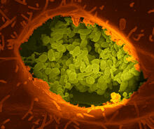
Q fever bacteria in an infected cell
3621
This image shows Q fever bacteria (yellow), which infect cows, sheep, and goats around the world and can infect humans, as well. When caught early, Q fever can be cured with antibiotics. Robert Heinzen, Elizabeth Fischer, and Anita Mora, National Institute of Allergy and Infectious Diseases, National Institutes of Health View Media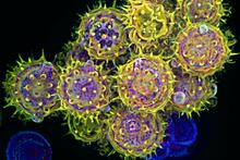
Pollen grains: male germ cells in plants and a cause of seasonal allergies
3609
Those of us who get sneezy and itchy-eyed every spring or fall may have pollen grains, like those shown here, to blame. Edna, Gil, and Amit Cukierman, Fox Chase Cancer Center, Philadelphia, Pa. View Media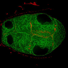
Fruit fly egg chamber
6811
A fruit fly (Drosophila melanogaster) egg chamber with microtubules shown in green and actin filaments shown in red. Vladimir I. Gelfand, Feinberg School of Medicine, Northwestern University. View Media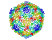
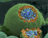
Bacteriophage P22 capsid
5874
Cryo-electron microscopy (cryo-EM) has the power to capture details of proteins and other small biological structures at the molecular level. This image shows proteins in the capsid, or outer co Dr. Wah Chiu, Baylor College of Medicine View Media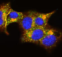
Insulin and protein interact in pancreatic beta cells
3546
A large number of proteins interact with the hormone insulin as it is produced in and secreted from the beta cells of the pancreas. William E. Balch, The Scripps Research Institute View Media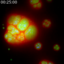
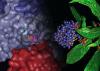
Nucleolus subcompartments spontaneously self-assemble 1
3789
The nucleolus is a small but very important protein complex located in the cell's nucleus. Nilesh Vaidya, Princeton University View Media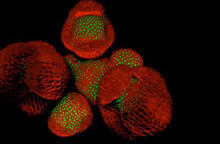
Arabidopsis Thaliana: Flowers Spring to Life
6503
This image capture shows how a single gene, STM, plays a starring role in plant development. Nathanaёl Prunet NIH Support: National Institute of General Medical Sciences View Media