Switch to Gallery View
Image and Video Gallery
This is a searchable collection of scientific photos, illustrations, and videos. The images and videos in this gallery are licensed under Creative Commons Attribution Non-Commercial ShareAlike 3.0. This license lets you remix, tweak, and build upon this work non-commercially, as long as you credit and license your new creations under identical terms.
Nuclear Lamina – Three Views
6573
Three views of the entire nuclear lamina of a HeLa cell produced by tilted light sheet 3D single-molecule super-resolution imaging using a platform termed TILT3D. Anna-Karin Gustavsson, Ph.D. View Media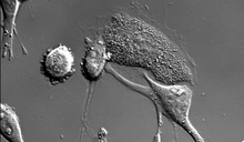
Dying melanoma cells
6966
Melanoma (skin cancer) cells undergoing programmed cell death, also called apoptosis. This process was triggered by raising the pH of the medium that the cells were growing in. Dylan T. Burnette, Vanderbilt University School of Medicine. View Media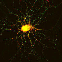
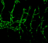
Neuron with labeled synapses
3509
In this image, recombinant probes known as FingRs (Fibronectin Intrabodies Generated by mRNA display) were expressed in a cortical neuron, where they attached fluorescent proteins to either PSD95 (gre Don Arnold and Richard Roberts, University of Southern California. View Media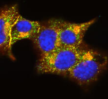
Insulin and protein interact in pancreatic beta cells
3546
A large number of proteins interact with the hormone insulin as it is produced in and secreted from the beta cells of the pancreas. William E. Balch, The Scripps Research Institute View Media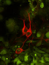
Three neurons and human ES cells
3290
The three neurons (red) visible in this image were derived from human embryonic stem cells. Undifferentiated stem cells are green here. Anirvan Ghosh lab, University of California, San Diego, via CIRM View Media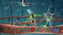
Motion in the brain
2323
Amid a network of blood vessels and star-shaped support cells, neurons in the brain signal each other. The mists of color show the flow of important molecules like glucose and oxygen. Kim Hager and Neal Prakash, University of California, Los Angeles View Media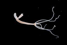
Hydra 05
2441
Hydra magnipapillata is an invertebrate animal used as a model organism to study developmental questions, for example the formation of the body axis. Hiroshi Shimizu, National Institute of Genetics in Mishima, Japan View Media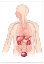
Body toxins
2496
Body organs such as the liver and kidneys process chemicals and toxins. These "target" organs are susceptible to damage caused by these substances. Crabtree + Company View Media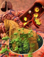
Host infection stimulates antibiotic resistance
5764
This illustration shows pathogenic bacteria behave like a Trojan horse: switching from antibiotic susceptibility to resistance during infection. View Media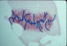
Lily mitosis 08
1021
A light microscope image of a cell from the endosperm of an African globe lily (Scadoxus katherinae). This is one frame of a time-lapse sequence that shows cell division in action. Andrew S. Bajer, University of Oregon, Eugene View Media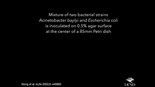
Time-lapse video of floral pattern in a mixture of two bacterial species, Acinetobacter baylyi and Escherichia coli, grown on a semi-solid agar for 24 hours
6550
This time-lapse video shows the emergence of a flower-like pattern in a mixture of two bacterial species, motile Acinetobacter baylyi and non-motile Escherichia coli (green), that are gr L. Xiong et al, eLife 2020;9: e48885 View Media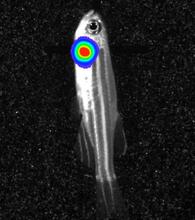
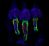
Bioluminescent imaging in adult zebrafish - lateral view
3558
Luciferase-based imaging enables visualization and quantification of internal organs and transplanted cells in live adult zebrafish. Kenneth Poss, Duke University View Media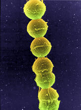
Streptococcus bacteria
1157
Image of Streptococcus, a type (genus) of spherical bacteria that can colonize the throat and back of the mouth. Stroptococci often occur in pairs or in chains, as shown here. Tina Weatherby Carvalho, University of Hawaii at Manoa View Media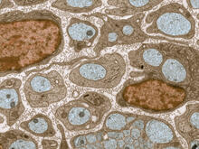
Transmission electron microscopy of myelinated axons with ECM between the axons
3736
The extracellular matrix (ECM) is most prevalent in connective tissues but also is present between the stems (axons) of nerve cells, as shown here. Tom Deerinck, National Center for Microscopy and Imaging Research (NCMIR) View Media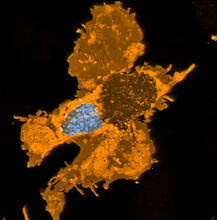
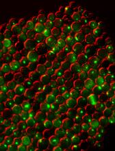
Zebrafish pigment cell
5754
Pigment cells are cells that give skin its color. David Parichy, University of Washington View Media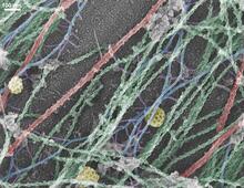
Cells use bubble-like structures called vesicles to transport cargo
3634
Cells use bubble-like structures called vesicles (yellow) to import, transport, and export cargo and in cellular communication. A single cell may be filled with thousands of moving vesicles.Tatyana Svitkina, University of Pennsylvania View Media
Protein clumping in zinc-deficient yeast cells
3550
The green spots in this image are clumps of protein inside yeast cells that are deficient in both zinc and a protein called Tsa1 that prevents clumping. Colin MacDiarmid and David Eide, University of Wisconsin--Madison View Media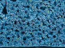
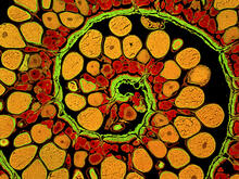
Anglerfish ovary cross-section
3620
This image captures the spiral-shaped ovary of an anglerfish in cross-section. Once matured, these eggs will be released in a gelatinous, floating mass. James E. Hayden, The Wistar Institute, Philadelphia, Pa. View Media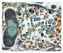
Cross section of a Drosophila melanogaster pupa lacking Draper
2759
In the absence of the engulfment receptor Draper, salivary gland cells (light blue) persist in the thorax of a developing Drosophila melanogaster pupa. Christina McPhee and Eric Baehrecke, University of Massachusetts Medical School View Media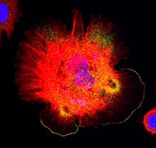
Spreading Cells 01
3328
Cells move forward with lamellipodia and filopodia supported by networks and bundles of actin filaments. Proper, controlled cell movement is a complex process. Rong Li and Praveen Suraneni, Stowers Institute for Medical Research View Media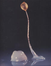
Dicty fruit
2684
Dictyostelium discoideum is a microscopic amoeba. A group of 100,000 form a mound as big as a grain of sand. Featured in The New Genetics. View Media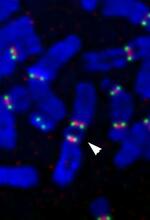
Fused, dicentric chromosomes
2763
This fused chromosome has two functional centromeres, shown as two sets of red and green dots. Beth A. Sullivan, Duke University View Media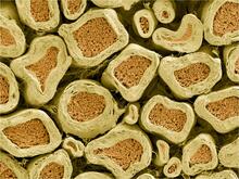
Myelinated axons 2
3397
Top view of myelinated axons in a rat spinal root. Tom Deerinck, National Center for Microscopy and Imaging Research (NCMIR) View Media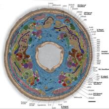
Annotated TEM cross-section of C. elegans (roundworm)
5760
The worm Caenorhabditis elegans is a popular laboratory animal because its small size and fairly simple body make it easy to study. Piali Sengupta, Brandeis University View Media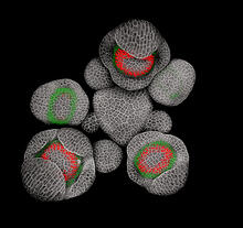
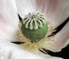
Developing Arabidopsis flower buds
3743
Flower development is a carefully orchestrated, genetically programmed process that ensures that the male (stamen) and female (pistil) organs form in the right place and at the right time in the flowe Nathanaël Prunet, Caltech View Media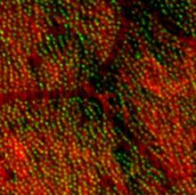
Centrioles anchor cilia in planaria
3292
Centrioles (green) anchor cilia (red), which project on the surface of pharynx cells of the freshwater planarian Schmidtea mediterranea. Juliette Azimzadeh, University of California, San Francisco View Media
Blinking bacteria
2724
Like a pulsing blue shower, E. coli cells flash in synchrony. Genes inserted into each cell turn a fluorescent protein on and off at regular intervals. Jeff Hasty, University of California, San Diego View Media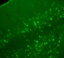
Confocal microscopy of perineuronal nets in the brain 2
3742
The photo shows a confocal microscopy image of perineuronal nets (PNNs), which are specialized extracellular matrix (ECM) structures in the brain. Tom Deerinck, National Center for Microscopy and Imaging Research (NCMIR) View Media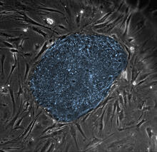
Human embryonic stem cells
2608
The center cluster of cells, colored blue, shows a colony of human embryonic stem cells. James Thomson, University of Wisconsin-Madison View Media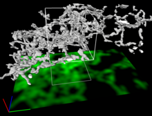
Dense tubular matrices in the peripheral endoplasmic reticulum (ER) 2
5856
Three-dimensional reconstruction of a tubular matrix in a thin section of the peripheral endoplasmic reticulum between the plasma membranes of the cell. Jennifer Lippincott-Schwartz, Howard Hughes Medical Institute Janelia Research Campus, Virginia View Media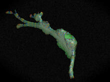
Cell curvature
2803
Rendering of the surface of an endothelial cell; membrane curvature is color coded. This is an example of NIH-supported research on single-cell analysis. Gaudenz Danuser, Harvard Medical School View Media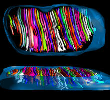
Mitochondrion from insect flight muscle
3662
This is a tomographic reconstruction of a mitochondrion from an insect flight muscle. National Center for Microscopy and Imaging Research View Media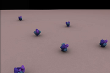
Clathrin-mediated endocytosis
5753
Endocytosis is the process by which cells are able to take up membrane and extracellular materials through the formation of a small intracellular bubble, called a vesicle. Janet Iwasa, University of Utah View Media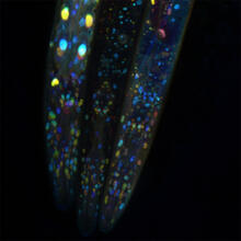
Mosaicism in C. elegans (Black Background)
6532
In the worm C. elegans, double-stranded RNA made in neurons can silence matching genes in a variety of cell types through the transport of RNA between cells. Snusha Ravikumar, Ph.D., University of Maryland, College Park, and Antony M. Jose, Ph.D., University of Maryland, College Park View Media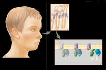
Olfactory system
1291
Sensory organs have cells equipped for detecting signals from the environment, such as odors. Judith Stoffer View Media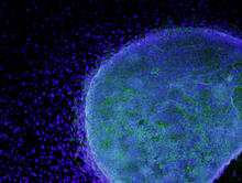
Colony of human ES cells
3269
A colony of human embryonic stem cells (light blue) grows on fibroblasts (dark blue). California Institute for Regenerative Medicine View Media
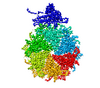
Bacteriophage P22 capsid, detail
5875
Detail of a subunit of the capsid, or outer cover, of bacteriophage P22, a virus that infects the Salmonella bacteria. Dr. Wah Chiu, Baylor College of Medicine View Media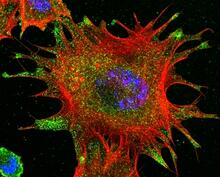
mDia1 antibody staining- 02
3331
Cells move forward with lamellipodia and filopodia supported by networks and bundles of actin filaments. Proper, controlled cell movement is a complex process. Rong Li and Praveen Suraneni, Stowers Institute for Medical Research View Media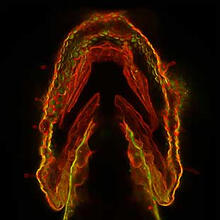
Glowing glycans
2473
Sugars light up the cells in this jaw of a 3-day-old zebrafish embryo and highlight a scientific first: labeling and tracking the movements of sugar chains called glycans in a living organism. Carolyn Bertozzi, University of California, Berkeley View Media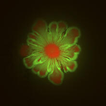
Floral pattern in a mixture of two bacterial species, Acinetobacter baylyi and Escherichia coli, grown on a semi-solid agar for 48 hours (photo 1)
6553
Floral pattern emerging as two bacterial species, motile Acinetobacter baylyi (red) and non-motile Escherichia coli (green), are grown together for 48 hours on 1% agar surface from a sma L. Xiong et al, eLife 2020;9: e48885 View Media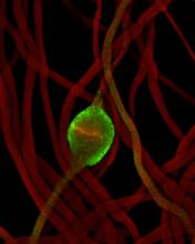
Fruit fly sperm cells
2433
Developing fruit fly spermatids require caspase activity (green) for the elimination of unwanted organelles and cytoplasm via apoptosis. Hermann Steller, Rockefeller University View Media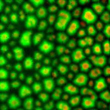
Cell-like compartments from frog eggs 3
6586
Cell-like compartments that spontaneously emerged from scrambled frog eggs. Endoplasmic reticulum (red) and microtubules (green) are visible. Image created using epifluorescence microscopy. Xianrui Cheng, Stanford University School of Medicine. View Media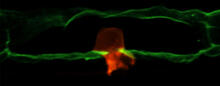
Anchor cell in basement membrane
2707
An anchor cell (red) pushes through the basement membrane (green) that surrounds it. Elliott Hagedorn, Duke University. View Media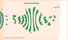
Cisternae maturation model
1307
Animation for the cisternae maturation model of Golgi transport. Judith Stoffer View Media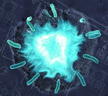
Supernova bacteria
2725
Bacteria engineered to act as genetic clocks flash in synchrony. Here, a "supernova" burst in a colony of coupled genetic clocks just after reaching critical cell density. Jeff Hasty, UCSD View Media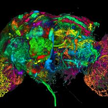
Color coding of the Drosophila brain - black background
5868
This image results from a research project to visualize which regions of the adult fruit fly (Drosophila) brain derive from each neural stem cell. Yong Wan from Charles Hansen’s lab, University of Utah. Data preparation and visualization by Masayoshi Ito in the lab of Kei Ito, University of Tokyo. View Media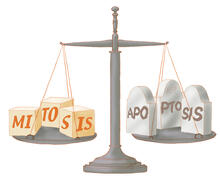
Life in balance
1336
Mitosis creates cells, and apoptosis kills them. The processes often work together to keep us healthy. Judith Stoffer View Media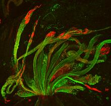
Fruit fly spermatids
3590
Developing spermatids (precursors of mature sperm cells) begin as small, round cells and mature into long-tailed, tadpole-shaped ones. Lacramioara Fabian, The Hospital for Sick Children, Toronto, Canada View Media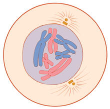
Mitosis - prophase
1330
A cell in prophase, near the start of mitosis: In the nucleus, chromosomes condense and become visible. In the cytoplasm, the spindle forms. Judith Stoffer View Media