Most cells are naturally
colorless, which is why scientists often use fluorescent tags and other tools
to color cell structures and make them easier to study. (Check out the
Pathways imaging issue
for more on scientific imaging techniques). Here, we’re showcasing cell images
that feature shades of blue. Visit our Image and Video Gallery
for additional images of cells in all the colors of the rainbow, as well as
other scientific photos, illustrations, and videos.
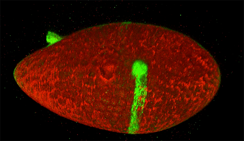
Credit: Jasmin Imran Alsous and Jonathan Jackson, Martin Lab, Massachusetts Institute of Technology.
What looks like a bubbling lava lamp is actually part of an egg cell’s maturation process. In many animals, the egg cell develops alongside sister cells. These sister cells are called nurse cells in the fruit fly (Drosophila melanogaster), and their job is to “nurse” an immature egg cell, or oocyte. Toward the end of oocyte development, the nurse cells transfer all their contents into the oocyte in a process called nurse cell dumping. This video captures this transfer, showing significant shape changes on the part of the nurse cells (blue), which are powered by wavelike activity of the protein myosin (red).
This post is a great supplement to Pathways: The Imaging Issue.
The video was taken using a confocal laser scanning microscopy (sometimes shortened to just “confocal microscopy”), one of the techniques mentioned in the Pathways timeline (1970s).
Have you ever wondered what creates striking images of cells and other tiny
structures? Most often, the answer is microscopes. Many of us have encountered
basic light microscopes in science classes, but those are just one of many
types that scientists use. Check out the slideshow to see images researchers
have captured using different kinds of microscopes. For even more images of
the microscopic world, visit the
NIGMS Image and Video Gallery.
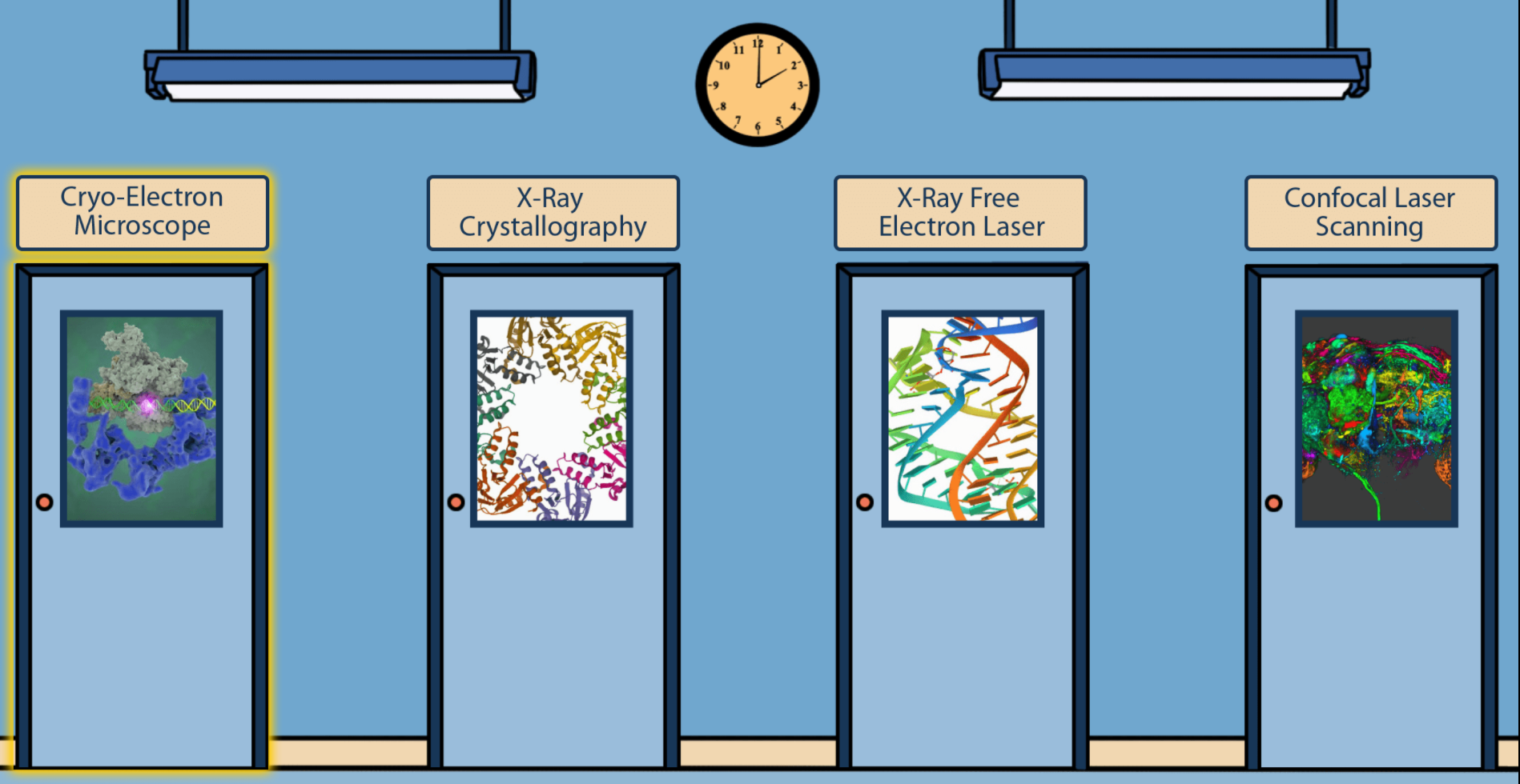
Students, teachers, and other curious minds can step into a scientific imaging lab with a free online interactive developed by NIGMS and Scholastic. Imaging tools help scientists unlock the mysteries of our cells and molecules. A better understanding of this tiny world can help researchers learn about the body’s normal and abnormal processes and lead to more effective, targeted treatments for illnesses.
NIGMS and Scholastic bring you our latest issue of Pathways, which focuses on imaging tools that help scientists unlock the mysteries of our cells and molecules. A better understanding of this tiny world can help researchers learn about the body’s normal and abnormal processes and lead to more effective, targeted treatments for illnesses.
Pathways is designed for students in grades 6 through 12. This collection of free resources teaches students about basic science and its importance to health, as well as exciting research careers.
To get a look at cell components that are too small to see with a normal light microscope, scientists often use cryo-electron microscopy (cryo-EM). As the prefix cryo- means “cold” or “freezing,” cryo-EM involves rapidly freezing a cell, virus, molecular complex, or other structure to prevent water molecules from forming crystals. This preserves the sample in its natural state and keeps it still so that it can be imaged with an electron microscope, which uses beams of electrons instead of light. Some electrons are scattered by the sample, while others pass through it and through magnetic lenses to land on a detector and form an image.
Typically, samples contain many copies of the object a scientist wants to study, frozen in a range of orientations. Researchers take images of these various positions and combine them into a detailed 3D model of the structure. Electron microscopes allow us to see much smaller structures than light microscopes do because the wavelengths of electrons are much shorter than the wavelength of light. NIGMS-funded researchers are using cryo-EM to investigate a range of scientific questions.
Caught in Translation
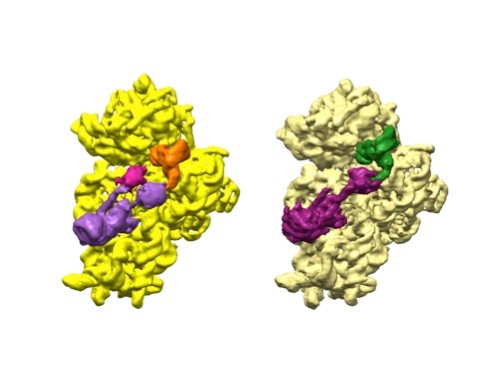
3D reconstructions of two stages in the assembly of the bacterial ribosome created from time-resolved cryo-EM images. Credit: Joachim Frank, Columbia University.
Joachim Frank, Ph.D., a professor of biochemistry and molecular biophysics and of biological sciences at Columbia University in New York, New York, along with two other researchers, won the 2017 Nobel Prize in Chemistry for developing cryo.
Dr. Frank’s lab focuses on the process of translation, where structures called ribosomes turn genetic instructions into proteins, which are needed for many chemical reactions that support life. Recently, Dr. Frank has adopted and further developed a technique called time-resolved cryo-EM. This method captures images of short-lived states in translation that disappear too quickly (after less than a second) for standard cryo-EM to capture. The ability to fully visualize translation could help researchers identify errors in the process that lead to disease and also to develop treatments.
Transformations aren’t just for people or pets around Halloween. Scientific images also can look different than you might expect, depending on how they’re photographed. Check out these tricky-looking images and learn more about the science behind them.
Credit: Nilay Taneja, Vanderbilt University, and Dylan T. Burnette, Ph.D., Vanderbilt University School of Medicine.
Do you have a hunch about what this image is? Perhaps something to do with dry leaves? It’s a human fibroblast cell undergoing cell division, or cytokinesis, into two daughter cells. Cytokinesis is essential for the growth and development of new cells. And fibroblasts play a big role in wound healing by helping with contraction and closure.
Microtubules sprout from one another. Credit: Petry lab, Princeton University.
The red spray pictured here may look like fireworks erupting across the night sky on July 4th, but it’s actually a rare glimpse of tiny protein strands called microtubules sprouting and growing from one another in a lab. Microtubules are the largest of the molecules that form a cell’s skeleton. When a cell divides, microtubules help ensure that each daughter cell has a complete set of genetic information from the parent. They also help organize the cell’s interior and even act as miniature highways for certain proteins to travel along.
As their name suggests, microtubules are hollow tubes made of building blocks called tubulins. Scientists know that a protein called XMAP215 adds tubulin proteins to the ends of microtubules to make them grow, but until recently, the way that a new microtubule starts forming remained a mystery.
Sabine Petry and her colleagues at Princeton University developed a new imaging method for watching microtubules as they develop and found an important clue to the mystery. They adapted a technique called total internal reflection fluorescence (TIRF) microscopy, which lit up only a tiny sliver of a sample from frog egg (Xenopus) tissue. This allowed the scientists to focus clearly on a few of the thousands of microtubules in a normal cell. They could then see what happened when they added certain proteins to the sample.
An engineered cell (green) in a fruit fly follicle (red), or egg case, leaves a trail of fluorescent material as it moves across a fruit fly egg chamber, allowing scientists to trace its path and measure how long it took to complete its journey. Credit: David Bilder, University of California, Berkeley.
Cells are the basis of the living world. Our cells make up the tissues and organs of our bodies. Bacteria are also cells, living sometimes alone and sometimes in groups called biofilms. We think of cells mostly as staying in one spot, quietly doing their work. But in many situations, cells move, often very quickly. For example, when you get a cut, infection-fighting cells rally to the site, ready to gobble up bacterial intruders. Then, platelet cells along with proteins from blood gather and form a clot to stop any bleeding. And finally, skin cells surrounding the wound lay down scaffolding before gliding across the cut to close the wound.
This remarkable organization and timing is evident right from the start. Cells migrate within the embryo as it develops so that body tissues and organs end up in the right places. Harmful cells use movement as well, as when cells move and spread (metastasize) from an original cancer tumor to other parts of the body. Learning how and why cells move could give scientists new ways to guide those cells or turn off or slow down the movement when needed.
Glowing Breadcrumbs
Scientists studying how humans and animals form, from a single cell at conception to a complex body at birth, are particularly interested in how and when cells move. They use research organisms like the fruit fly, Drosophila, to watch movements by small populations of cells. Still, watching cells migrate inside a living fly is challenging because the tissue is too dense to see individual cell movement. But moving those cells to a dish in the lab might cause them to behave differently than they do inside the fly. To solve this problem, NIGMS-funded researcher David Bilder and colleagues at the University of California, Berkeley, came up with a way to alter fly cells so they could track how the cells behave without removing them from the fly. They engineered the cells to lay down a glowing track of proteins behind them as they moved, leaving a traceable path through the fly’s tissue. The technique, called M-TRAIL (matrix-labeling technique for real-time and inferred location), allows the researchers to see where a cell travels and how long it takes to get there.
Bilder and his team first used M-TRAIL in flies to confirm the results of past studies of Drosophila ovaries in the lab using other imaging techniques. In addition, they found that M-TRAIL could be used to study a variety of cell types. The new technique also could allow a cell’s movement to be tracked over a longer period than other imaging techniques, which become toxic to cells in just a few hours. This is important, because cells often migrate for days to reach their final destinations.
The yellow-green glow from this summer’s fireflies teased my kids across the yard. Max and Stella zigzagged the grass, occasionally jumping into the air to cup a firefly in their hands and then proudly shouting, “I got one!”
Chasing fireflies on a summer night is a childhood rite of passage for many, including Nathan Shaner who grew up in New Jersey. “It was one of my favorite things about summer,” he recalls. “I’d catch them with my hands—I’d never jar them.”
Today, Shaner studies the science of bioluminescence, which gives fireflies and many other organisms the natural ability to emit light. His goal is to make bright bioluminescent tags that he and other scientists can use to study living cells in greater detail. “There’s this very beautiful thing that evolved in nature, and we can use it to enable new discoveries,” he says.
Thousands of organisms glow as a way to communicate, spook predators, lure prey or attract mates. There are a few terrestrial examples, such as fireflies, glowworm insect larvae and foxfire fungi, and many more aquatic ones, including types of marine plankton, fish, jellyfish, shrimp, squid and sea urchins. One research team estimated nearly three quarters of sea life have bioluminescent capabilities.

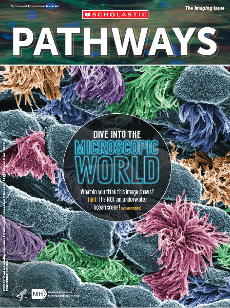 Cover of Pathways student magazine.
 3D reconstructions of two stages in the assembly of the bacterial ribosome created from time-resolved cryo-EM images. Credit: Joachim Frank, Columbia University.
 Credit: Nilay Taneja, Vanderbilt University, and Dylan T. Burnette, Ph.D., Vanderbilt University School of Medicine.
 Firefly. Credit: Stock photo.