Category: Cells

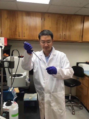
Career Conversations: Q&A With Biologist Akhila Rajan

November 3, 2021
A headshot of Dr. Akhila Rajan. Dr. Akhila Rajan. Credit: Fred Hutchinson Cancer Research Center.

“What makes being a scientist exciting is that I don’t know what I’m going to find tomorrow,” says Akhila Rajan, Ph.D., an assistant professor in the basic sciences division at Fred Hutchinson Cancer Research Center in Seattle, Washington. Dr. Rajan is supported by an NIGMS early stage investigator Maximizing Investigators’ Research Award. These awards provide stable and flexible funding for a program of research that falls within NIGMS’ mission. Check out the highlights of our interview with Dr. Rajan to learn about her research and journey as a scientist.

Continue Reading

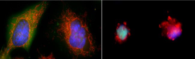
Cool Images: Spooky and Spectacular

October 27, 2021

This time of year, many creepy crawlies take center stage to frighten people of all ages. To celebrate Halloween, we've conjured up a slideshow of fascinating but spooky species that NIGMS-funded scientists study. Some of these creatures drink blood like vampires, while others—frogs, worms, flies, and salamanders—are perfect cauldron ingredients for a witch's brew. Check out the slideshow—if you dare!

For other less frightening images, visit our image and video gallery to find scientific photos, illustrations, and videos.

Continue Reading

Science Snippet: Brush Up on Biofilms

September 22, 2021

A biofilm is a highly organized community of microorganisms that develops naturally on certain surfaces. Typically, biofilms are made up of microbes and an extracellular matrix that they produce. This matrix can include polysaccharides (chains of sugars), proteins, lipids, DNA, and other molecules. The matrix gives the biofilm structure and helps it stick to a surface.

Formation of a biofilm often involves a process called quorum sensing. In this process, microbes detect when they reach a certain population density and change their behavior in ways that help them function as a community.

Continue Reading

Make Like a Cell and Split: Comparing Mitosis and Meiosis

September 8, 2021

Your body is made up of trillions of cells that all originate from just one—a fertilized egg. The massive multiplication of cells after conception is possible thanks to cell division, which occurs when one cell splits into two. Cell division not only enables growth but also replaces damaged or dead cells and makes reproduction possible. There are two kinds of cell division: mitosis and meiosis.

On the left, a cell goes through the stages of mitosis to split into two cells that each have two sets of chromosomes. On the right, a cell goes through the phases of meiosis to divide into four cells that each have a single set of chromosomes. 
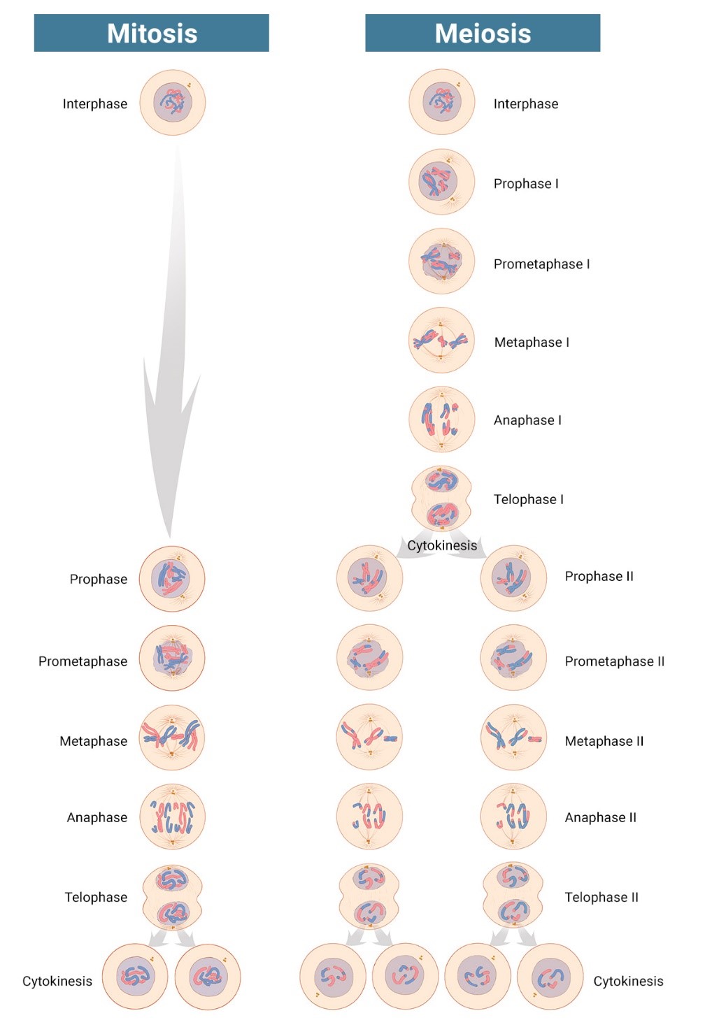
Mitosis is shown on the left, and meiosis is shown on the right. Credit: Judith Stoffer. Click to enlarge
Continue Reading

Cool Images: Beautiful Bits of Blue

August 18, 2021

Most cells are naturally colorless, which is why scientists often use fluorescent tags and other tools to color cell structures and make them easier to study. (Check out the Pathways imaging issue for more on scientific imaging techniques). Here, we’re showcasing cell images that feature shades of blue. Visit our Image and Video Gallery for additional images of cells in all the colors of the rainbow, as well as other scientific photos, illustrations, and videos.

Continue Reading

Science Snippet: Apoptosis Explained

July 28, 2021

Apoptosis is the process by which cells in the body die in a controlled and predictable way because they have DNA damage or are no longer needed. The term comes from a Greek word meaning “falling off,” as in leaves falling from a tree.

When a cell undergoes apoptosis, it shrinks and pulls away from its neighbors. As the cytoskeleton that gives it shape and structure collapses, the envelope around the cell’s nucleus breaks down, and its DNA breaks into pieces. Its surface changes, signaling its death to other cells and leading a healthy cell to engulf the dying one and recycle its components.

On the left, two large cells with clear, smooth edges. On the right, two smaller cells with ragged edges.
Two cells in a healthy state (left) and entering apoptosis (right). Credit: Hogan Tang of the Denise Montell Lab, Johns Hopkins School of Medicine.
Continue Reading

Cool Video: A Biological Lava Lamp

July 21, 2021
Several spheres contorting and lighting up inside a cone-shaped structure.
Credit: Jasmin Imran Alsous and Jonathan Jackson, Martin Lab, Massachusetts Institute of Technology.

What looks like a bubbling lava lamp is actually part of an egg cell’s maturation process. In many animals, the egg cell develops alongside sister cells. These sister cells are called nurse cells in the fruit fly (Drosophila melanogaster), and their job is to “nurse” an immature egg cell, or oocyte. Toward the end of oocyte development, the nurse cells transfer all their contents into the oocyte in a process called nurse cell dumping. This video captures this transfer, showing significant shape changes on the part of the nurse cells (blue), which are powered by wavelike activity of the protein myosin (red).

Researchers created the video using a confocal laser scanning microscope. Learn about this type of microscope and other scientific imaging tools by stepping into our virtual imaging lab, and check out more basic science videos and photos in the NIGMS Image and Video Gallery.

This post is a great supplement to Pathways: The Imaging Issue.

The video was taken using a confocal laser scanning microscopy (sometimes shortened to just “confocal microscopy”), one of the techniques mentioned in the Pathways timeline (1970s).

Learn more in our Educator's Corner.

Continue Reading

Career Conversations: Q&A with Molecular Biologist Hong Liu

July 7, 2021
A scientist wearing a lab coat and holding a pipette in front of a workbench with scientific instruments.
Dr. Hong Liu in the lab.
Credit: Courtesy of Dr. Hong Liu.

“A scientific career is really worth it,” says Hong Liu, Ph.D., an assistant professor of biochemistry and molecular biology at Tulane University School of Medicine in New Orleans, Louisiana. Check out the highlights of our interview with Dr. Liu below to learn about his journey as a scientist and his advice for students.

Q: What makes a career in science exciting?

A: I think there are at least two things that make a science career very exciting. The first is that doing science means you have freedom to explore a lot of new ideas. The second thing is it’s rewarding. The "rewarding" I’m talking about here is not like how much money you can make. It’s rewarding in the answers you find and the new knowledge you reveal.

Continue Reading

A Focus on Microscopes: See Eye-Catching Images

June 30, 2021

Have you ever wondered what creates striking images of cells and other tiny structures? Most often, the answer is microscopes. Many of us have encountered basic light microscopes in science classes, but those are just one of many types that scientists use. Check out the slideshow to see images researchers have captured using different kinds of microscopes. For even more images of the microscopic world, visit the NIGMS Image and Video Gallery.

Continue Reading

Career Conversations: Q&A with Microbiologist Josephine Chandler

June 9, 2021

Josephine (Josie) Chandler, Ph.D., first became interested in science when she took a high school chemistry class. In college, she fell in love with microbiology and ultimately earned a Ph.D. in the field. Today, she’s an associate professor of molecular biosciences at the University of Kansas in Lawrence, where her lab investigates interactions in bacterial communities. By better understanding these interactions, scientists may find new ways to stop infections or break down environmental pollutants—a process known as bioremediation.

Continue Reading