If you’ve ever visited an aquarium or snorkeled along a coral reef, you’ve witnessed the dazzling colors and patterns on tropical fish. The iridescent stripes and dots come from pigment cells, which also tint skin, hair and eyes in all kinds of animals, including humans. Typically, bright colors help attract mates, while dull ones provide camouflage. In humans, pigment helps protect skin from DNA-damaging UV light.
Researchers study cellular hues not only to decipher how they color our world, but also to understand skin cancers that originate from pigment cells. Some of these researchers work their way back, developmentally speaking, to focus on the type of cell, known as a neural crest cell, that is the precursor of pigment cells.
Present at the earliest stages of development, neural crest cells migrate throughout an embryo and transform into many different types of cells and tissues, including nerve cells, cartilage, bone and skin. The images here, from research on neural crest cells in fish and salamanders, showcase the beauty and versatility of pigment cells in nature’s palette.
Pigment cells called xanthophores, shown here in the skin of the popular laboratory animal zebrafish, glow brightly under light. Credit: David Parichy, University of Washington.
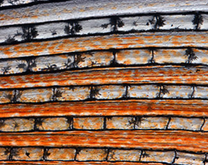
Dark pigment cells, called melanocytes, like these in pearl danio, a tropical minnow and relative of zebrafish, assemble in skin patterns that allow the animals to blend into their surroundings or attract mates. Credit: David Parichy, University of Washington.
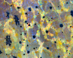
Pigment cells can form all sorts of patterns, like these stripes on the fin of pearl danio. Credit: David Parichy, University of Washington.
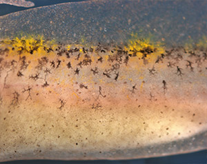
Pigment cells arise from neural crest cells. Here, pigment cells can be seen migrating in the skin of a salamander where they will form distinct color patterns. Credit: David Parichy, University of Washington.
Hard labor might be the very thing we try to avoid on Labor Day. But our cells and their components don’t have the luxury of taking a day off. Their non-stop work is what keeps us going and healthy.
Scientists often compare cells with small factories. Just like a factory, a cell contains specialized compartments and machines—including organelles and other structures—that each play their own roles in getting the job done. In the vignettes below, we give a shout out to some of these tireless cellular workers.
Energy Generators
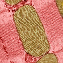
Mitochondria are the cell’s power plants. They convert energy from food into a molecule called ATP that fuels virtually every process in the cell. As shown here, mitochondria (brown) often have distinct, oblong shapes. Like most other organelles, mitochondria are encased in an outer membrane. But they also have an inner membrane that folds many times, increasing the area available for energy production. In addition, mitochondria store calcium ions, help make hemoglobin—the vital iron-containing protein that allows red blood cells to carry oxygen—and even take part in producing some hormones. Defects in mitochondria can lead to a host of rare but often incurable diseases that range from mild to devastating. Researchers are studying mitochondria to better understand their manifold jobs in the cell and to find treatments for mitochondrial diseases.
The world beneath our skin is full of movement. Hemoglobin in our blood grabs oxygen and delivers it throughout the body. Molecular motors in cells chug along tiny tubes, hauling cargo with them. Biological invaders like viruses enter our bodies, hijack our cells and reproduce wildly before bursting out to infect other cells.
To make sense of the subcutaneous world, Janet Iwasa, a molecular animator at the University of Utah, creates “visual hypotheses”—detailed animations that convey the latest thinking of how biological molecules interact.
“It’s really building the animated model that brings insights,” Iwasa told Biomedical Beat in 2014. “When you’re creating an animation, you’re really grappling with a lot of issues that don’t necessarily come up by any other means. In some cases, it might raise more questions, and make people go back and do some more experiments when they realize there might be something missing.”
Iwasa has collaborated with numerous scientists to develop animations of a range of biological processes and structures. Recently, she’s undertaken an ambitious, multi-year project to animate HIV reproduction.
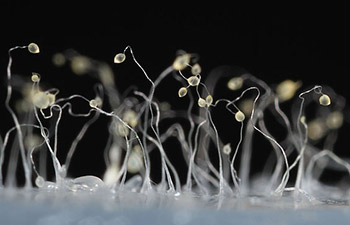
Dictyostelium discoideumNatural habitat: Deciduous forest soil and moist leaf litter
Favorite food: Bacteria
Top speed: 8 micrometers per minute
Like the athletes in Rio, the world’s most highly advanced microbial runners recently gathered in Charlestown, Massachusetts, to find out which ones could use chemical cues to most quickly navigate a maze-like microfluidic racecourse. The winners’ prize: credit for helping scientists learn more about how immune system cells navigate through the human body on their way to fight disease.
The finalists were a group of soil-dwelling slime molds called Dictyostelium that were genetically engineered by a pair of Dutch biochemists to detect minuscule chemical changes in the environment. The racers used their enhanced sense of “smell” to avoid getting lost on their way to the finish line.
While researchers have been racing the genetically souped-up microbes at annual events for a few years—another competition is scheduled for October 26—scientists have been studying conventional Dictyostelium for decades to investigate other important basic life processes including early development, gene function, self/non-self recognition, cell-type regulation, chemical signaling and programmed cell death.
Enrique M. De La CruzGrew up in: Newark and Kearny, New Jersey
Job site: Yale University
Favorite food: His mom’s Spanish-style polenta (harina de maíz)
Alternative career: Managing a vinyl record shop
Favorite song: “Do Anything You Wanna Do” by Eddie & The Hot Rods
Enrique De La Cruz stood off to the side in a packed room. As he waited for his turn to speak, he stroked the beads of a necklace. Was he nervous? Quietly praying? When he took center stage, the purpose of the strand became clear.
Like a magician—and dressed all in black—De La Cruz held up the necklace with two hands so everyone, even those sitting in the back, could see it. It was made of snap-together beads. De La Cruz waved the strand. It wiggled in different directions. Then, with no sleight of hand, he popped off one of the beads. The necklace broke into two.
For the next hour, De La Cruz pulled out one prop after another: a piece of rope from his pocket, a pencil tucked behind his ear and even a fresh spear of asparagus stuffed in his backpack. At one point, De La Cruz assembled a conga line with people in the front row.
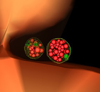
This illustration of the inside of a yeast cell shows two mature, or “late” endosomes (green-ringed structures) that are filled with small vesicles (red bubbles). Endosomes are cellular containers that can carry many types of cargo, including cellular waste, which they typically dump into vacuoles (orange). Credit: Matthew West and Greg Odorizzi, University of Colorado, Boulder.
In large offices, mailroom workers read the labels on incoming letters and packages to sort and deliver them and dispose of junk mail. In cells, these tasks—as well as importing food and other materials—fall to small cellular sacs called endosomes. Acting as mailroom staff, endosomes sort and deliver nutrients and building blocks, like amino acids, fat and sugars, to their proper destinations, and send cellular junk, like damaged proteins, to trash processors, such as vacuoles or lysosomes.
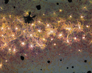
Researchers created an apparatus to study quorum sensing, a communication system that allows some bacteria to cause dangerous infections. Their findings suggest that blocking bacterial communication might lead to a new way to combat such infections. Credit: Minyoung Kevin Kim and Bonnie Bassler, Princeton University.
If you’ve ever felt a slimy coating on your teeth, scrubbed grime from around a sink drain or noticed something growing between the tiles of a shower, you’ve encountered a biofilm. Made up of communities of bacteria and other microorganisms, biofilms thrive where they can remain moist and relatively undisturbed. As they enlarge, biofilms can block narrow passages like medical stents, airways, pipes or intestines.
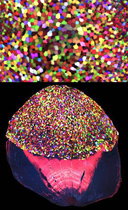
More than 70 Skinbow colors distinguish hundreds of live cells from a tiny bit (0.0003348 square inches) of skin on the tail fin of an adult zebrafish. The bottom image shows the cells on the outer surface of a scale. Credit: Chen-Hui Chen, Duke University.
Zebrafish, blue-and-white-striped fish that are about 1.5 inches long, can regrow injured or lost fins. This feature makes the small fish a useful model organism for scientists who study tissue regeneration.
To better understand how zebrafish skin recovers after a scrape or amputation, researchers led by Kenneth Poss of Duke University tracked thousands of skin cells in real time. They found that lifespans of individual skin cells on the surface were 8 to 9 days on average and that the entire skin surface turned over in 20 days.
The scientists used an imaging technique they developed called “Skinbow,” which essentially shows the fish’s outer layer of skin cells in a spectrum of colors when viewed under a microscope. Skinbow is based on a technique created to study nerve cells in mice, another model organism.
The research team’s color-coded experiments revealed several unexpected cellular responses during tissue repair and replacement. The scientists plan to incorporate additional imaging techniques to generate an even more detailed picture of the tissue regeneration process.
The NIH director showcased the Skinbow technique and these images on his blog, writing: “You can see more than 70 detectable Skinbow colors that make individual cells as visually distinct from one another as jellybeans in a jar.”
This work was funded in part by NIH under grant R01GM074057.
Our cells are constantly removing and recycling molecular waste. On the occasion of Earth Day, we put together this narrated animation to show you one way cells process their trash. The video features the proteasome, a cellular machine that breaks down damaged or unwanted proteins into bits that the cell can re-use to make new proteins. For this reason, the proteasome is as much a recycling plant as it is a garbage disposal.
For more details about the proteasome and other cellular disposal systems, check out our article How Cells Take Out the Trash.
Studying some of the most well-tread territory in science can turn up surprising new findings. Take, for example, the cell. You may have read in textbooks how the cell’s parts look and function during important biological processes like cellular movement and division. You may have even built models of the cell out of gelatin or clay. But scientists continue to learn new facts that require those textbooks to be updated, and those models to be reshaped. Here are a few examples.
Nuclear Envelope: More Than a Protective Barrier
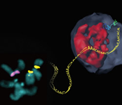
Damaged heterochromatin, a tightly packed form of DNA, travels to the inner wall of the nuclear envelope for repair. Credit: Irene Chiolo and Taehyun Ryu, University of Southern California.
Like a security guard checking IDs at the door, the nuclear envelope forms a protective barrier around the cell’s nucleus, only letting specific proteins and chemical signals pass through. Scientists recently found that this envelope may also act as a repair center for broken strands of heterochromatin, a tightly packed form of DNA.
Irene Chiolo of the University of Southern California and Gary Karpen of the University of California, Berkeley, and the Lawrence Berkeley National Laboratory were part of a team that learned that healthy fruit fly cells mend breaks in heterochromatin by moving the damaged DNA strands to the inner wall of the nuclear envelope. There, proteins embedded in the envelope make the necessary repairs in a safe place where the broken DNA can’t accidentally get fused to the wrong chromosome.

 Credit: Wikimedia Commons, Usman Bashir.
 Credit: Jeff Foley, American Heart Association.